- PDB-5d0r: Crystal structure of human soluble Adenylyl Cyclase with the inhi... -
+
Open data
ID or keywords:
Loading...
-
Basic information
Entry
Database: PDB / ID: 5d0r
Title
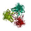
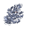
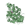
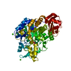
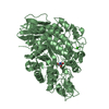
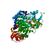
Crystal structure of human soluble Adenylyl Cyclase with the inhibitor bithionol
Components
Adenylate cyclase type 10
Keywords
LYASE
Function / homology
Function and homology information
negative regulation of cardiac muscle cell contraction / mitochondrial ATP transmembrane transport / bicarbonate binding / neuron projection retraction / epithelial cilium movement involved in extracellular fluid movement / astrocyte end-foot / central region of growth cone / glucose catabolic process / positive regulation of glycogen catabolic process / regulation of mitophagy ...negative regulation of cardiac muscle cell contraction / mitochondrial ATP transmembrane transport / bicarbonate binding / neuron projection retraction / epithelial cilium movement involved in extracellular fluid movement / astrocyte end-foot / central region of growth cone / glucose catabolic process / positive regulation of glycogen catabolic process / regulation of mitophagy / regulation of membrane repolarization / adenylate cyclase / positive regulation of oxidative stress-induced neuron intrinsic apoptotic signaling pathway / basal part of cell / adenylate cyclase activity / positive regulation of ossification / cAMP biosynthetic process / neuron projection extension / : / positive regulation of vascular associated smooth muscle cell apoptotic process / positive regulation of cardiac muscle hypertrophy / positive regulation of reactive oxygen species biosynthetic process / positive regulation of mitochondrial depolarization / negative regulation of mitochondrial membrane potential / positive regulation of ATP biosynthetic process / spermatid development / positive regulation of axon extension / Hedgehog 'off' state / negative regulation of reactive oxygen species biosynthetic process / neuron projection maintenance / positive regulation of cardiac muscle cell apoptotic process / apical part of cell / adenylate cyclase-inhibiting G protein-coupled receptor signaling pathway / manganese ion binding / ATPase binding / cytoskeleton / intracellular signal transduction / cilium / neuronal cell body / dendrite / perinuclear region of cytoplasm / magnesium ion binding / mitochondrion / extracellular region / ATP binding / nucleus / plasma membrane / cytoplasm / cytosol Similarity search - Function
Resolution: 2.24→86.23 Å / Cor.coef. Fo:Fc: 0.958 / Cor.coef. Fo:Fc free: 0.912 / SU B: 14.431 / SU ML: 0.177 / Cross valid method: THROUGHOUT / σ(F): 0 / ESU R: 0.256 / ESU R Free: 0.218 / Stereochemistry target values: MAXIMUM LIKELIHOOD Details: HYDROGENS HAVE BEEN ADDED IN THE RIDING POSITIONS U VALUES : WITH TLS ADDED
Rfactor
Num. reflection
% reflection
Selection details
Rfree
0.2437
1383
5.1 %
RANDOM
Rwork
0.1773
-
-
-
obs
0.1806
25668
99.74 %
-
Solvent computation
Ion probe radii: 0.8 Å / Shrinkage radii: 0.8 Å / VDW probe radii: 1.2 Å / Solvent model: MASK
In the structure databanks used in Yorodumi, some data are registered as the other names, "COVID-19 virus" and "2019-nCoV". Here are the details of the virus and the list of structure data.
Jan 31, 2019. EMDB accession codes are about to change! (news from PDBe EMDB page)
EMDB accession codes are about to change! (news from PDBe EMDB page)
The allocation of 4 digits for EMDB accession codes will soon come to an end. Whilst these codes will remain in use, new EMDB accession codes will include an additional digit and will expand incrementally as the available range of codes is exhausted. The current 4-digit format prefixed with “EMD-” (i.e. EMD-XXXX) will advance to a 5-digit format (i.e. EMD-XXXXX), and so on. It is currently estimated that the 4-digit codes will be depleted around Spring 2019, at which point the 5-digit format will come into force.
The EM Navigator/Yorodumi systems omit the EMD- prefix.
Related info.:Q: What is EMD? / ID/Accession-code notation in Yorodumi/EM Navigator
Yorodumi is a browser for structure data from EMDB, PDB, SASBDB, etc.
This page is also the successor to EM Navigator detail page, and also detail information page/front-end page for Omokage search.
The word "yorodu" (or yorozu) is an old Japanese word meaning "ten thousand". "mi" (miru) is to see.
Related info.:EMDB / PDB / SASBDB / Comparison of 3 databanks / Yorodumi Search / Aug 31, 2016. New EM Navigator & Yorodumi / Yorodumi Papers / Jmol/JSmol / Function and homology information / Changes in new EM Navigator and Yorodumi
 Movie
Movie Controller

 Yorodumi
 Open data
 Basic information
 Components
 Keywords
 Function and homology information
 Homo sapiens (human)
 X-RAY DIFFRACTION /
  Authors
 Germany, 1items
  Citation
 Structure visualization
 Downloads & links
 Other downloads
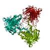


 PDBj
PDBj


 Assembly
 Trichoplusia ni (cabbage looper) / References: UniProt: Q96PN6, adenylate cyclase


 Mass: 356.052 Da / Num. of mol.: 1 / Source method: obtained synthetically / Formula: C12H6Cl4O2S / Comment: antibiotic*YM
 Mass: 59.044 Da / Num. of mol.: 1 / Source method: obtained synthetically / Formula: C2H3O2
 Mass: 62.068 Da / Num. of mol.: 6 / Source method: obtained synthetically / Formula: C2H6O2
 Mass: 78.133 Da / Num. of mol.: 3 / Source method: obtained synthetically / Formula: C2H6OS / Comment: DMSO, precipitant*YM
 Mass: 92.094 Da / Num. of mol.: 2 / Source method: obtained synthetically / Formula: C3H8O3
 Sample preparation
 Processing