 Movie
Movie Controller

[English] 日本語
 Yorodumi
- PDB-1zja: Crystal structure of the trehalulose synthase MutB from Pseudomon... -
+ Open data

- Basic information
| Entry | Database: PDB / ID: 1zja | ||||||
|---|---|---|---|---|---|---|---|
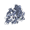
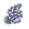
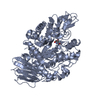
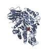
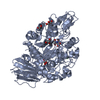
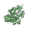
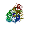
| Title | Crystal structure of the trehalulose synthase MutB from Pseudomonas mesoacidophila MX-45 (triclinic form) | ||||||
 Components Components | Trehalulose synthase | ||||||
 Keywords Keywords | ISOMERASE / trehalulose synthase / sucrose isomerase / alpha-amylase family / (beta/alpha)8 barrel | ||||||
| Function / homology |  Function and homology information Function and homology informationalpha-amylase activity / oligosaccharide catabolic process / isomerase activity / metal ion binding Similarity search - Function | ||||||
| Biological species |  Pseudomonas mesoacidophila (bacteria) Pseudomonas mesoacidophila (bacteria) | ||||||
| Method |  X-RAY DIFFRACTION / SYNCHROTRON / MOLECULAR REPLACEMENT / Resolution: 1.6 Å X-RAY DIFFRACTION / SYNCHROTRON / MOLECULAR REPLACEMENT / Resolution: 1.6 Å | ||||||
 Authors Authors | Ravaud, S. / Robert, X. / Haser, R. / Aghajari, N. | ||||||
 Citation Citation | Journal: J.Biol.Chem. / Year: 2007 Title: Trehalulose synthase native and carbohydrate complexed structures provide insights into sucrose isomerization. Authors: Ravaud, S. / Robert, X. / Watzlawick, H. / Haser, R. / Mattes, R. / Aghajari, N. #1: Journal: Acta Crystallogr.,Sect.F / Year: 2005Title: Expression, purification, crystallization and preliminary X-ray crystallographic studies of the trehalulose synthase MutB from Pseudomonas mesoacidophila MX-45. Authors: Ravaud, S. / Watzlawick, H. / Haser, R. / Mattes, R. / Aghajari, N. | ||||||
| History |
| ||||||
| Remark 999 | SEQUENCE According to authors, there is no database reference for the protein sequence as it is ... SEQUENCE According to authors, there is no database reference for the protein sequence as it is patented (U.S. Patent *5786140*). |
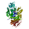
- Structure visualization
| Structure viewer | Molecule: MolmilJmol/JSmol |
|---|
- Downloads & links
-Download
| PDBx/mmCIF format | 1zja.cif.gz | 273.3 KB | Display | PDBx/mmCIF format |
|---|---|---|---|---|
| PDB format | pdb1zja.ent.gz | 216.2 KB | Display | PDB format |
| PDBx/mmJSON format | 1zja.json.gz | Tree view | PDBx/mmJSON format | |
| Others |  Other downloads Other downloads |
-Validation report
| Arichive directory | https://data.pdbj.org/pub/pdb/validation_reports/zj/1zjaftp://data.pdbj.org/pub/pdb/validation_reports/zj/1zja | HTTPS FTP |
|---|
-Related structure data
| Related structure data | 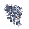 2pwdC 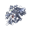 2pweC 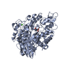 2pwfC 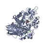 2pwgC 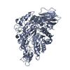 2pwhC  1m53S S: Starting model for refinement C: citing same article ( |
|---|---|
| Similar structure data |


-Links
 PDBj
PDBj

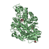
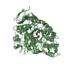
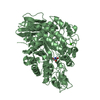
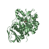
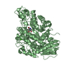
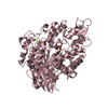
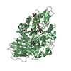
- Assembly
| Deposited unit | 
| ||||||||
|---|---|---|---|---|---|---|---|---|---|
| 1 | 
| ||||||||
| 2 | 
| ||||||||
| Unit cell |
|
-Components
| #1: Protein | Mass: 63938.848 Da / Num. of mol.: 2 Source method: isolated from a genetically manipulated source Source: (gene. exp.) Pseudomonas mesoacidophila (bacteria) / Strain: MX-45 / Plasmid: pJOE2702 / Production host: #2: Chemical |   Mass: 40.078 Da / Num. of mol.: 2 / Source method: obtained synthetically / Formula: Ca Mass: 40.078 Da / Num. of mol.: 2 / Source method: obtained synthetically / Formula: Ca#3: Chemical |   Mass: 122.143 Da / Num. of mol.: 2 / Source method: obtained synthetically / Formula: C4H12NO3 / Comment: pH buffer*YM Mass: 122.143 Da / Num. of mol.: 2 / Source method: obtained synthetically / Formula: C4H12NO3 / Comment: pH buffer*YM#4: Water | ChemComp-HOH / |  Mass: 18.015 Da / Num. of mol.: 1703 / Source method: isolated from a natural source / Formula: H2O Mass: 18.015 Da / Num. of mol.: 1703 / Source method: isolated from a natural source / Formula: H2O |
|---|
-Experimental details
-Experiment
| Experiment | Method: X-RAY DIFFRACTION / Number of used crystals: 1 |
|---|
- Sample preparation
| Crystal | Density Matthews: 2.5 Å3/Da / Density % sol: 51 % |
|---|---|
| Crystal grow | Temperature: 290 K / Method: vapor diffusion, hanging drop / pH: 8.5 Details: PEG 20000, Tris-HCl, L-cysteine, pH 8.5, temperature 290K, VAPOR DIFFUSION, HANGING DROP |
-Data collection
| Diffraction | Mean temperature: 100 K |
|---|---|
| Diffraction source | Source: SYNCHROTRON / Site: ESRF  / Beamline: ID14-4 / Wavelength: 1.072 Å / Beamline: ID14-4 / Wavelength: 1.072 Å |
| Detector | Type: ADSC QUANTUM 4 / Detector: CCD / Date: May 10, 2004 |
| Radiation | Monochromator: synchrotron optics / Protocol: SINGLE WAVELENGTH / Monochromatic (M) / Laue (L): M / Scattering type: x-ray |
| Radiation wavelength | Wavelength: 1.072 Å / Relative weight: 1 |
| Reflection | Resolution: 1.6→39 Å / Num. all: 156700 / Num. obs: 156700 / % possible obs: 95 % / Observed criterion σ(F): 1 / Observed criterion σ(I): 1 / Redundancy: 1.9 % / Biso Wilson estimate: 15.2 Å2 / Rsym value: 0.086 / Net I/σ(I): 4.7 |
| Reflection shell | Resolution: 1.6→1.7 Å / Redundancy: 2 % / Mean I/σ(I) obs: 2.2 / Rsym value: 0.277 / % possible all: 95 |
- Processing
| Software |
| ||||||||||||||||||||||||||||||||||||
|---|---|---|---|---|---|---|---|---|---|---|---|---|---|---|---|---|---|---|---|---|---|---|---|---|---|---|---|---|---|---|---|---|---|---|---|---|---|
| Refinement | Method to determine structure: MOLECULAR REPLACEMENT Starting model: PDB ENTRY 1M53 Resolution: 1.6→39 Å / Rfactor Rfree error: 0.002 / Data cutoff high absF: 930872.34 / Data cutoff low absF: 0 / Isotropic thermal model: RESTRAINED / Cross valid method: THROUGHOUT / σ(F): 0 / Stereochemistry target values: Engh & Huber
| ||||||||||||||||||||||||||||||||||||
| Solvent computation | Solvent model: FLAT MODEL / Bsol: 47.7909 Å2 / ksol: 0.367044 e/Å3 | ||||||||||||||||||||||||||||||||||||
| Displacement parameters | Biso mean: 13.9 Å2
| ||||||||||||||||||||||||||||||||||||
| Refine analyze |
| ||||||||||||||||||||||||||||||||||||
| Refinement step | Cycle: LAST / Resolution: 1.6→39 Å
| ||||||||||||||||||||||||||||||||||||
| Refine LS restraints |
| ||||||||||||||||||||||||||||||||||||
| LS refinement shell | Resolution: 1.6→1.7 Å / Rfactor Rfree error: 0.006 / Total num. of bins used: 6
| ||||||||||||||||||||||||||||||||||||
| Xplor file |
|
