 Movie
Movie Controller

[English] 日本語
 Yorodumi
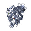
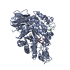
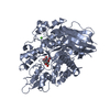
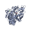
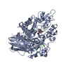
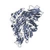
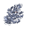
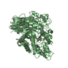
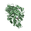
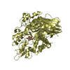
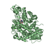
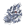
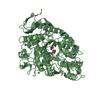
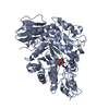
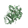
- PDB-4gi6: Crystal structure of the MUTB F164L mutant in complex with glucose -
+ Open data

- Basic information
| Entry | Database: PDB / ID: 4gi6 | ||||||
|---|---|---|---|---|---|---|---|
| Title | Crystal structure of the MUTB F164L mutant in complex with glucose | ||||||
 Components Components | Sucrose isomerase | ||||||
 Keywords Keywords | ISOMERASE / Enzyme complex / Tim-barrel(beta/alpha)8 / Sucrose isomerase / Glycoside hydrolase / Trehalulose synthase / GH13 family(cazy database) / Calcium binding | ||||||
| Function / homology |  Function and homology information Function and homology informationalpha-amylase activity / oligosaccharide catabolic process / isomerase activity / metal ion binding Similarity search - Function | ||||||
| Biological species |  Rhizobium (bacteria) Rhizobium (bacteria) | ||||||
| Method |  X-RAY DIFFRACTION / SYNCHROTRON / FOURIER SYNTHESIS / Resolution: 2.15 Å X-RAY DIFFRACTION / SYNCHROTRON / FOURIER SYNTHESIS / Resolution: 2.15 Å | ||||||
 Authors Authors | Lipski, A. / Haser, R. / Aghajari, N. | ||||||
 Citation Citation | Journal: Acta Crystallogr.,Sect.D / Year: 2013 Title: Mutations inducing an active-site aperture in Rhizobium sp. sucrose isomerase confer hydrolytic activity Authors: Lipski, A. / Watzlawick, H. / Ravaud, S. / Robert, X. / Rhimi, M. / Haser, R. / Mattes, R. / Aghajari, N. | ||||||
| History |
|
- Structure visualization
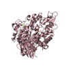
| Structure viewer | Molecule: MolmilJmol/JSmol |
|---|
- Downloads & links
-Download
| PDBx/mmCIF format | 4gi6.cif.gz | 252.6 KB | Display | PDBx/mmCIF format |
|---|---|---|---|---|
| PDB format | pdb4gi6.ent.gz | 200.7 KB | Display | PDB format |
| PDBx/mmJSON format | 4gi6.json.gz | Tree view | PDBx/mmJSON format | |
| Others |  Other downloads Other downloads |
-Validation report
| Arichive directory | https://data.pdbj.org/pub/pdb/validation_reports/gi/4gi6ftp://data.pdbj.org/pub/pdb/validation_reports/gi/4gi6 | HTTPS FTP |
|---|
-Related structure data
| Related structure data | 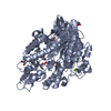 4gi8C 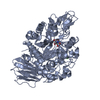 4gi9C 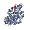 4giaC  4ginC 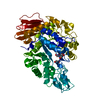 4h2cC 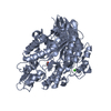 1zjbS C: citing same article ( S: Starting model for refinement |
|---|---|
| Similar structure data |


-Links
 PDBj
PDBj


- Assembly
| Deposited unit | 
| ||||||||
|---|---|---|---|---|---|---|---|---|---|
| 1 | 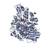
| ||||||||
| 2 | 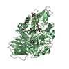
| ||||||||
| Unit cell |
|
-Components
-Protein / Sugars , 2 types, 3 molecules AB
| #1: Protein | Mass: 63904.832 Da / Num. of mol.: 2 / Fragment: MUTB fragment, UNP residues 28-584 / Mutation: F164L Source method: isolated from a genetically manipulated source Source: (gene. exp.) Rhizobium (bacteria) / Strain: MX-45 / Gene: mutB / Plasmid: PHWG660.12 / Production host: References: UniProt: Q2PS28, UniProt: M1E1F3*PLUS, EC: 5.4.11.99 #3: Sugar | ChemComp-GLC / |  Type: D-saccharide, alpha linking / Mass: 180.156 Da / Num. of mol.: 1 Type: D-saccharide, alpha linking / Mass: 180.156 Da / Num. of mol.: 1Source method: isolated from a genetically manipulated source Formula: C6H12O6 |
|---|
-Non-polymers , 4 types, 903 molecules 


| #2: Chemical |  Mass: 40.078 Da / Num. of mol.: 2 / Source method: obtained synthetically / Formula: Ca Mass: 40.078 Da / Num. of mol.: 2 / Source method: obtained synthetically / Formula: Ca#4: Chemical | ChemComp-GOL /  Mass: 92.094 Da / Num. of mol.: 8 / Source method: obtained synthetically / Formula: C3H8O3 Mass: 92.094 Da / Num. of mol.: 8 / Source method: obtained synthetically / Formula: C3H8O3#5: Chemical | ChemComp-TRS / |  Mass: 122.143 Da / Num. of mol.: 1 / Source method: obtained synthetically / Formula: C4H12NO3 / Comment: pH buffer*YM Mass: 122.143 Da / Num. of mol.: 1 / Source method: obtained synthetically / Formula: C4H12NO3 / Comment: pH buffer*YM#6: Water | ChemComp-HOH / | Mass: 18.015 Da / Num. of mol.: 892 / Source method: isolated from a natural source / Formula: H2O |
|---|
-Experimental details
-Experiment
| Experiment | Method: X-RAY DIFFRACTION / Number of used crystals: 1 |
|---|
- Sample preparation
| Crystal | Density Matthews: 2.53 Å3/Da / Density % sol: 51.35 % |
|---|---|
| Crystal grow | Temperature: 292 K / Method: vapor diffusion, hanging drop / pH: 8 Details: 0.1M Tris-HCl, 12%(w/v) PEG20000, 0.01M L-Cysteine, pH 8, VAPOR DIFFUSION, HANGING DROP, temperature 292K |
-Data collection
| Diffraction | Mean temperature: 100 K |
|---|---|
| Diffraction source | Source: SYNCHROTRON / Site: ESRF  / Beamline: ID29 / Wavelength: 0.97559 Å / Beamline: ID29 / Wavelength: 0.97559 Å |
| Detector | Type: ADSC QUANTUM 315r / Detector: CCD / Date: Apr 11, 2008 |
| Radiation | Monochromator: YALE MIRRORS / Protocol: SINGLE WAVELENGTH / Monochromatic (M) / Laue (L): M / Scattering type: x-ray |
| Radiation wavelength | Wavelength: 0.97559 Å / Relative weight: 1 |
| Reflection | Resolution: 2.15→15 Å / Num. all: 69351 / Num. obs: 68858 / % possible obs: 99.3 % / Observed criterion σ(F): 0 / Observed criterion σ(I): 1 / Redundancy: 3.7 % / Biso Wilson estimate: 26.2 Å2 / Rsym value: 0.117 / Net I/σ(I): 9.4 |
| Reflection shell | Resolution: 2.15→2.3 Å / Redundancy: 3.5 % / Rmerge(I) obs: 0.464 / Mean I/σ(I) obs: 3.3 / Num. unique all: 12519 / % possible all: 99.4 |
- Processing
| Software |
| |||||||||||||||||||||||||||||||||||||||||||||
|---|---|---|---|---|---|---|---|---|---|---|---|---|---|---|---|---|---|---|---|---|---|---|---|---|---|---|---|---|---|---|---|---|---|---|---|---|---|---|---|---|---|---|---|---|---|---|
| Refinement | Method to determine structure: FOURIER SYNTHESIS Starting model: 1ZJB Resolution: 2.15→14.91 Å / Cor.coef. Fo:Fc: 0.968 / Cor.coef. Fo:Fc free: 0.943 / SU B: 4.89 / SU ML: 0.126 / Cross valid method: THROUGHOUT / σ(F): 2 / ESU R: 0.232 / ESU R Free: 0.188 / Stereochemistry target values: MAXIMUM LIKELIHOOD
| |||||||||||||||||||||||||||||||||||||||||||||
| Solvent computation | Ion probe radii: 0.8 Å / Shrinkage radii: 0.8 Å / VDW probe radii: 1.2 Å / Solvent model: MASK | |||||||||||||||||||||||||||||||||||||||||||||
| Displacement parameters | Biso mean: 19.31 Å2
| |||||||||||||||||||||||||||||||||||||||||||||
| Refinement step | Cycle: LAST / Resolution: 2.15→14.91 Å
| |||||||||||||||||||||||||||||||||||||||||||||
| Refine LS restraints |
| |||||||||||||||||||||||||||||||||||||||||||||
| LS refinement shell | Resolution: 2.15→2.206 Å / Total num. of bins used: 20
|
