 Movie
Movie Controller

[English] 日本語
 Yorodumi
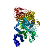
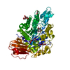
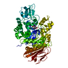
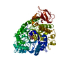
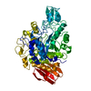
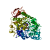
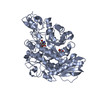
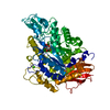
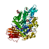
- PDB-1m53: CRYSTAL STRUCTURE OF ISOMALTULOSE SYNTHASE (PALI) FROM KLEBSIELLA... -
+ Open data

- Basic information
| Entry | Database: PDB / ID: 1m53 | ||||||
|---|---|---|---|---|---|---|---|
| Title | CRYSTAL STRUCTURE OF ISOMALTULOSE SYNTHASE (PALI) FROM KLEBSIELLA SP. LX3 | ||||||
 Components Components | Isomaltulose Synthase | ||||||
 Keywords Keywords | ISOMERASE / KLEBSIELLA SP. LX3 / ISOMALTULOSE SYNTHASE / SUCROSE ISOMERIZATION | ||||||
| Function / homology |  Function and homology information Function and homology informationisomaltulose synthase / alpha-amylase activity / oligosaccharide catabolic process Similarity search - Function | ||||||
| Biological species |  Klebsiella sp. LX3 (bacteria) Klebsiella sp. LX3 (bacteria) | ||||||
| Method |  X-RAY DIFFRACTION / SYNCHROTRON / MOLECULAR REPLACEMENT / Resolution: 2.2 Å X-RAY DIFFRACTION / SYNCHROTRON / MOLECULAR REPLACEMENT / Resolution: 2.2 Å | ||||||
 Authors Authors | Li, N. / Swaminathan, K. | ||||||
 Citation Citation | Journal: J.Biol.Chem. / Year: 2003 Title: Isomaltulose synthase (PalI) of Klebsiella sp. LX3. Crystal structure and implication of mechanism Authors: Zhang, D. / Li, N. / Lok, S.M. / Zhang, L.-H. / Swaminathan, K. #1: Journal: APPL.ENVIRON.MICROBIOL. / Year: 2002Title: Isomaltulose Synthase from Klebsiella sp. Strain LX3: Gene Cloning and Characterization and Engineering of Thermostability Authors: Zhang, D.-H. / Li, X.-Z. / Zhang, L.-H. | ||||||
| History |
|
- Structure visualization
| Structure viewer | Molecule: MolmilJmol/JSmol |
|---|
- Downloads & links
-Download
| PDBx/mmCIF format | 1m53.cif.gz | 133.9 KB | Display | PDBx/mmCIF format |
|---|---|---|---|---|
| PDB format | pdb1m53.ent.gz | 103.2 KB | Display | PDB format |
| PDBx/mmJSON format | 1m53.json.gz | Tree view | PDBx/mmJSON format | |
| Others |  Other downloads Other downloads |
-Validation report
| Arichive directory | https://data.pdbj.org/pub/pdb/validation_reports/m5/1m53ftp://data.pdbj.org/pub/pdb/validation_reports/m5/1m53 | HTTPS FTP |
|---|
-Related structure data
| Related structure data |  1uokS S: Starting model for refinement |
|---|---|
| Similar structure data |
-Links
 PDBj
PDBj


- Assembly
| Deposited unit | 
| ||||||||
|---|---|---|---|---|---|---|---|---|---|
| 1 |
| ||||||||
| Unit cell |
|
-Components
| #1: Protein | Mass: 67251.797 Da / Num. of mol.: 1 / Fragment: residues 29-598 Source method: isolated from a genetically manipulated source Source: (gene. exp.) Klebsiella sp. LX3 (bacteria) / Gene: PalI / Plasmid: pGEX-2T / Species (production host): Escherichia coli / Production host: |
|---|---|
| #2: Water | ChemComp-HOH /  Mass: 18.015 Da / Num. of mol.: 303 / Source method: isolated from a natural source / Formula: H2O Mass: 18.015 Da / Num. of mol.: 303 / Source method: isolated from a natural source / Formula: H2O |
-Experimental details
-Experiment
| Experiment | Method: X-RAY DIFFRACTION / Number of used crystals: 1 |
|---|
- Sample preparation
| Crystal | Density Matthews: 2.31 Å3/Da / Density % sol: 46.34 % | |||||||||||||||||||||||||||||||||||
|---|---|---|---|---|---|---|---|---|---|---|---|---|---|---|---|---|---|---|---|---|---|---|---|---|---|---|---|---|---|---|---|---|---|---|---|---|
| Crystal grow | Temperature: 293 K / Method: vapor diffusion, hanging drop / pH: 6.5 Details: 0.1M SODIUM CACODYLATE, 0.2M AMMONIUM SULPHATE, 30%(W/V) PEG8000, pH 6.5, VAPOR DIFFUSION, HANGING DROP, temperature 293K | |||||||||||||||||||||||||||||||||||
| Crystal grow | *PLUS Temperature: 22 ℃ / pH: 7.5 / Method: vapor diffusion, hanging drop | |||||||||||||||||||||||||||||||||||
| Components of the solutions | *PLUS
|
-Data collection
| Diffraction | Mean temperature: 100 K |
|---|---|
| Diffraction source | Source: SYNCHROTRON / Site: SPring-8  / Beamline: BL40B2 / Wavelength: 1 / Wavelength: 1 Å / Beamline: BL40B2 / Wavelength: 1 / Wavelength: 1 Å |
| Detector | Type: ADSC QUANTUM 4 / Detector: CCD / Date: Apr 5, 2002 |
| Radiation | Monochromator: DIAMOND / Protocol: SINGLE WAVELENGTH / Monochromatic (M) / Laue (L): M / Scattering type: x-ray |
| Radiation wavelength | Wavelength: 1 Å / Relative weight: 1 |
| Reflection | Resolution: 2.2→20 Å / Num. obs: 32456 / % possible obs: 99.2 % / Observed criterion σ(F): 3 / Redundancy: 6.1 % / Biso Wilson estimate: 21.3 Å2 / Rsym value: 0.053 / Net I/σ(I): -3 |
| Reflection shell | Resolution: 2.2→2.28 Å / Redundancy: 4.3 % / Rsym value: 0.269 / % possible all: 96.6 |
| Reflection | *PLUS Num. obs: 29756 / Num. measured all: 199659 / Rmerge(I) obs: 0.053 |
| Reflection shell | *PLUS % possible obs: 96.6 % / Rmerge(I) obs: 0.26 |
- Processing
| Software |
| ||||||||||||||||||||
|---|---|---|---|---|---|---|---|---|---|---|---|---|---|---|---|---|---|---|---|---|---|
| Refinement | Method to determine structure: MOLECULAR REPLACEMENT Starting model: PDB CODE: 1UOK Resolution: 2.2→8 Å / Rfactor Rfree error: 0.004 / Data cutoff high absF: 264716.53 / Data cutoff low absF: 0 / Isotropic thermal model: RESTRAINED / Cross valid method: THROUGHOUT / σ(F): 3
| ||||||||||||||||||||
| Solvent computation | Solvent model: FLAT MODEL / Bsol: 51.9159 Å2 / ksol: 0.440529 e/Å3 | ||||||||||||||||||||
| Displacement parameters | Biso mean: 37.5 Å2
| ||||||||||||||||||||
| Refine analyze |
| ||||||||||||||||||||
| Refinement step | Cycle: LAST / Resolution: 2.2→8 Å
| ||||||||||||||||||||
| Refine LS restraints |
| ||||||||||||||||||||
| LS refinement shell | Resolution: 2.2→2.33 Å / Rfactor Rfree error: 0.013 / Total num. of bins used: 6
| ||||||||||||||||||||
| Xplor file |
| ||||||||||||||||||||
| Refinement | *PLUS Highest resolution: 2.2 Å / Lowest resolution: 20 Å | ||||||||||||||||||||
| Solvent computation | *PLUS | ||||||||||||||||||||
| Displacement parameters | *PLUS | ||||||||||||||||||||
| Refine LS restraints | *PLUS
| ||||||||||||||||||||
| LS refinement shell | *PLUS Lowest resolution: 2.28 Å |
