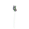
 Movie
Movie Controller

[English] 日本語
 Yorodumi
- PDB-5cti: Crystal structure of the type IX collagen NC2 hetero-trimerizatio... -
+ Open data

- Basic information
| Entry | Database: PDB / ID: 5cti | ||||||
|---|---|---|---|---|---|---|---|
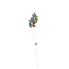
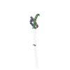
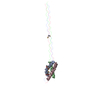
| Title | Crystal structure of the type IX collagen NC2 hetero-trimerization domain with a guest fragment a2a1a1 of type I collagen (native form) | ||||||
 Components Components |
| ||||||
 Keywords Keywords | STRUCTURAL PROTEIN / collagen / hetero-trimerization / chain stagger / chain register / triple helix | ||||||
| Function / homology |  Function and homology information Function and homology informationcollagen type IX trimer / collagen type I trimer / cellular response to vitamin E / tooth mineralization / protein heterotrimerization / cellular response to fluoride / Anchoring fibril formation / intramembranous ossification / Crosslinking of collagen fibrils / cellular response to acetaldehyde ...collagen type IX trimer / collagen type I trimer / cellular response to vitamin E / tooth mineralization / protein heterotrimerization / cellular response to fluoride / Anchoring fibril formation / intramembranous ossification / Crosslinking of collagen fibrils / cellular response to acetaldehyde / extracellular matrix assembly / collagen biosynthetic process / Defective VWF binding to collagen type I / Collagen chain trimerization / Enhanced cleavage of VWF variant by ADAMTS13 / Defective VWF cleavage by ADAMTS13 variant / platelet-derived growth factor binding / bone trabecula formation / extracellular matrix structural constituent conferring tensile strength / Enhanced binding of GP1BA variant to VWF multimer:collagen / Defective binding of VWF variant to GPIb:IX:V / cartilage development involved in endochondral bone morphogenesis / embryonic skeletal system development / skin morphogenesis / collagen metabolic process / Collagen biosynthesis and modifying enzymes / collagen-activated tyrosine kinase receptor signaling pathway / Fibronectin matrix formation / endochondral ossification / Signaling by PDGF / Platelet Adhesion to exposed collagen / NCAM1 interactions / collagen fibril organization / response to steroid hormone / face morphogenesis / Assembly of collagen fibrils and other multimeric structures / odontogenesis / MET activates PTK2 signaling / Scavenging by Class A Receptors / extracellular matrix structural constituent / Syndecan interactions / GP1b-IX-V activation signalling / Platelet Aggregation (Plug Formation) / blood vessel development / RUNX2 regulates osteoblast differentiation / bone mineralization / SMAD binding / Collagen degradation / Non-integrin membrane-ECM interactions / basement membrane / negative regulation of cell-substrate adhesion / response to hyperoxia / ECM proteoglycans / Integrin cell surface interactions / response to cAMP / cellular response to transforming growth factor beta stimulus / positive regulation of epithelial to mesenchymal transition / protein localization to nucleus / Rho protein signal transduction / GPVI-mediated activation cascade / cellular response to retinoic acid / cellular response to fibroblast growth factor stimulus / visual perception / transforming growth factor beta receptor signaling pathway / secretory granule / cellular response to epidermal growth factor stimulus / animal organ morphogenesis / Cell surface interactions at the vascular wall / Developmental Lineage of Pancreatic Ductal Cells / skeletal system development / response to hydrogen peroxide / cellular response to amino acid stimulus / cellular response to mechanical stimulus / cellular response to glucose stimulus / sensory perception of sound / SMAD2/SMAD3:SMAD4 heterotrimer regulates transcription / response to insulin / regulation of blood pressure / cellular response to tumor necrosis factor / Immunoregulatory interactions between a Lymphoid and a non-Lymphoid cell / osteoblast differentiation / response to estradiol / positive regulation of canonical Wnt signaling pathway / protein transport / carbohydrate binding / extracellular matrix / protease binding / Interleukin-4 and Interleukin-13 signaling / protein-macromolecule adaptor activity / positive regulation of cell migration / response to xenobiotic stimulus / endoplasmic reticulum lumen / positive regulation of DNA-templated transcription / protein homodimerization activity / : / extracellular exosome / extracellular region / metal ion binding / identical protein binding Similarity search - Function | ||||||
| Biological species |  Homo sapiens (human) Homo sapiens (human) | ||||||
| Method |  X-RAY DIFFRACTION / MOLECULAR REPLACEMENT / Resolution: 1.8994 Å X-RAY DIFFRACTION / MOLECULAR REPLACEMENT / Resolution: 1.8994 Å | ||||||
 Authors Authors | Boudko, S.P. / Bachinger, H.P. | ||||||
| Funding support |  United States, 1items United States, 1items
| ||||||
 Citation Citation | Journal: Sci Rep / Year: 2016 Title: Structural insight for chain selection and stagger control in collagen. Authors: Boudko, S.P. / Bachinger, H.P. | ||||||
| History |
|
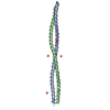
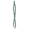
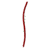
- Structure visualization
| Structure viewer | Molecule: MolmilJmol/JSmol |
|---|
- Downloads & links
-Download
| PDBx/mmCIF format | 5cti.cif.gz | 85.6 KB | Display | PDBx/mmCIF format |
|---|---|---|---|---|
| PDB format | pdb5cti.ent.gz | 65.3 KB | Display | PDB format |
| PDBx/mmJSON format | 5cti.json.gz | Tree view | PDBx/mmJSON format | |
| Others |  Other downloads Other downloads |
-Validation report
| Arichive directory | https://data.pdbj.org/pub/pdb/validation_reports/ct/5ctiftp://data.pdbj.org/pub/pdb/validation_reports/ct/5cti | HTTPS FTP |
|---|
-Related structure data
| Related structure data |  5ctdSC  5cvaC  5cvbC S: Starting model for refinement C: citing same article ( |
|---|---|
| Similar structure data |
-Links
 PDBj
PDBj


- Assembly
| Deposited unit | 
| ||||||||
|---|---|---|---|---|---|---|---|---|---|
| 1 |
| ||||||||
| Unit cell |
|
-Components
| #1: Protein | Mass: 7020.964 Da / Num. of mol.: 1 / Fragment: UNP Residues 572-583,UNP Residues 754-789 Source method: isolated from a genetically manipulated source Source: (gene. exp.) Homo sapiens (human) / Gene: COL1A1, COL9A1 / Plasmid: pET22b(+) / Production host:  | ||||
|---|---|---|---|---|---|
| #2: Protein | Mass: 6895.721 Da / Num. of mol.: 1 / Fragment: UNP Residues 484-495,UNP Residues 517-552 Source method: isolated from a genetically manipulated source Source: (gene. exp.) Homo sapiens (human) / Gene: COL1A2, COL9A2 / Plasmid: pET22b(+) / Production host: | ||||
| #3: Protein | Mass: 6999.000 Da / Num. of mol.: 1 / Fragment: UNP Residues 572-583,UNP Residues 517-553 Source method: isolated from a genetically manipulated source Source: (gene. exp.) Homo sapiens (human) / Gene: COL1A1, COL9A3 / Plasmid: pET22b(+) / Production host: | ||||
| #4: Chemical |   Mass: 92.094 Da / Num. of mol.: 3 / Source method: obtained synthetically / Formula: C3H8O3 Mass: 92.094 Da / Num. of mol.: 3 / Source method: obtained synthetically / Formula: C3H8O3#5: Water | ChemComp-HOH / |  Mass: 18.015 Da / Num. of mol.: 145 / Source method: isolated from a natural source / Formula: H2O Mass: 18.015 Da / Num. of mol.: 145 / Source method: isolated from a natural source / Formula: H2OHas protein modification | Y | |
-Experimental details
-Experiment
| Experiment | Method: X-RAY DIFFRACTION |
|---|
- Sample preparation
| Crystal | Density Matthews: 2.29 Å3/Da / Density % sol: 46.23 % |
|---|---|
| Crystal grow | Temperature: 295 K / Method: vapor diffusion, hanging drop / pH: 7.5 / Details: 0.1 M HEPES, 0.2 M sodium acetate, 17% PEG 3,350 |
-Data collection
| Diffraction | Mean temperature: 100 K | ||||||||||||||||||||||||||||||||||||||||||||||||||||||||||||||||||||||||||||||||||||||||||||||||||||||||||||||||||||||||||||||
|---|---|---|---|---|---|---|---|---|---|---|---|---|---|---|---|---|---|---|---|---|---|---|---|---|---|---|---|---|---|---|---|---|---|---|---|---|---|---|---|---|---|---|---|---|---|---|---|---|---|---|---|---|---|---|---|---|---|---|---|---|---|---|---|---|---|---|---|---|---|---|---|---|---|---|---|---|---|---|---|---|---|---|---|---|---|---|---|---|---|---|---|---|---|---|---|---|---|---|---|---|---|---|---|---|---|---|---|---|---|---|---|---|---|---|---|---|---|---|---|---|---|---|---|---|---|---|---|
| Diffraction source | Source: ROTATING ANODE / Type: RIGAKU MICROMAX-003 / Wavelength: 1.5418 Å | ||||||||||||||||||||||||||||||||||||||||||||||||||||||||||||||||||||||||||||||||||||||||||||||||||||||||||||||||||||||||||||||
| Detector | Type: RIGAKU SATURN 724 / Detector: CCD / Date: Jan 15, 2014 | ||||||||||||||||||||||||||||||||||||||||||||||||||||||||||||||||||||||||||||||||||||||||||||||||||||||||||||||||||||||||||||||
| Radiation | Protocol: SINGLE WAVELENGTH / Monochromatic (M) / Laue (L): M / Scattering type: x-ray | ||||||||||||||||||||||||||||||||||||||||||||||||||||||||||||||||||||||||||||||||||||||||||||||||||||||||||||||||||||||||||||||
| Radiation wavelength | Wavelength: 1.5418 Å / Relative weight: 1 | ||||||||||||||||||||||||||||||||||||||||||||||||||||||||||||||||||||||||||||||||||||||||||||||||||||||||||||||||||||||||||||||
| Reflection | Resolution: 1.8994→50 Å / Num. obs: 14864 / % possible obs: 99.1 % / Redundancy: 11.9 % / Rmerge(I) obs: 0.068 / Χ2: 1.607 / Net I/av σ(I): 40.585 / Net I/σ(I): 15.3 / Num. measured all: 176334 | ||||||||||||||||||||||||||||||||||||||||||||||||||||||||||||||||||||||||||||||||||||||||||||||||||||||||||||||||||||||||||||||
| Reflection shell | Diffraction-ID: 1 / Rejects: _
|
- Processing
| Software |
| ||||||||||||||||||||||||||||||||||||||||||||||||||||||||||||||||||||||||||||||||||||
|---|---|---|---|---|---|---|---|---|---|---|---|---|---|---|---|---|---|---|---|---|---|---|---|---|---|---|---|---|---|---|---|---|---|---|---|---|---|---|---|---|---|---|---|---|---|---|---|---|---|---|---|---|---|---|---|---|---|---|---|---|---|---|---|---|---|---|---|---|---|---|---|---|---|---|---|---|---|---|---|---|---|---|---|---|---|
| Refinement | Method to determine structure: MOLECULAR REPLACEMENT Starting model: 5CTD Resolution: 1.8994→30.804 Å / SU ML: 0.16 / Cross valid method: THROUGHOUT / σ(F): 1.34 / Phase error: 19.9 / Stereochemistry target values: ML
| ||||||||||||||||||||||||||||||||||||||||||||||||||||||||||||||||||||||||||||||||||||
| Solvent computation | Shrinkage radii: 0.9 Å / VDW probe radii: 1.11 Å / Solvent model: FLAT BULK SOLVENT MODEL | ||||||||||||||||||||||||||||||||||||||||||||||||||||||||||||||||||||||||||||||||||||
| Displacement parameters | Biso max: 84.42 Å2 / Biso mean: 26.6441 Å2 / Biso min: 6.69 Å2 | ||||||||||||||||||||||||||||||||||||||||||||||||||||||||||||||||||||||||||||||||||||
| Refinement step | Cycle: final / Resolution: 1.8994→30.804 Å
| ||||||||||||||||||||||||||||||||||||||||||||||||||||||||||||||||||||||||||||||||||||
| Refine LS restraints |
| ||||||||||||||||||||||||||||||||||||||||||||||||||||||||||||||||||||||||||||||||||||
| LS refinement shell | Refine-ID: X-RAY DIFFRACTION / Total num. of bins used: 11
|
