cornification / structural constituent of skin epidermis / keratin filament / Keratinization / positive regulation of epidermis development / complement activation, lectin pathway / intermediate filament organization / Formation of the cornified envelope / cornified envelope / Differentiation of Keratinocytes in Interfollicular Epidermis in Mammalian Skin ...cornification / structural constituent of skin epidermis / keratin filament / Keratinization / positive regulation of epidermis development / complement activation, lectin pathway / intermediate filament organization / Formation of the cornified envelope / cornified envelope / Differentiation of Keratinocytes in Interfollicular Epidermis in Mammalian Skin / intermediate filament / keratinization / protein heterotetramerization / regulation of angiogenesis / fibrinolysis / keratinocyte differentiation / morphogenesis of an epithelium / carbohydrate binding / signaling receptor activity / response to oxidative stress / blood microparticle / ficolin-1-rich granule lumen / cytoskeleton / protein heterodimerization activity / Neutrophil degranulation / cell surface / : / extracellular exosome / extracellular region / membrane / nucleus / cytoplasm / cytosol Similarity search - Function
Keratin type II cytoskeletal 1, tail / Keratin type II cytoskeletal 1 tail / Keratin, type II / Keratin type II head / Keratin type II head / Keratin, type I / Intermediate filament protein, conserved site / Intermediate filament protein / Intermediate filament (IF) rod domain signature. / Intermediate filament, rod domain ...Keratin type II cytoskeletal 1, tail / Keratin type II cytoskeletal 1 tail / Keratin, type II / Keratin type II head / Keratin type II head / Keratin, type I / Intermediate filament protein, conserved site / Intermediate filament protein / Intermediate filament (IF) rod domain signature. / Intermediate filament, rod domain / Intermediate filament (IF) rod domain profile. / Intermediate filament protein Similarity search - Domain/homology
In the structure databanks used in Yorodumi, some data are registered as the other names, "COVID-19 virus" and "2019-nCoV". Here are the details of the virus and the list of structure data.
Jan 31, 2019. EMDB accession codes are about to change! (news from PDBe EMDB page)
EMDB accession codes are about to change! (news from PDBe EMDB page)
The allocation of 4 digits for EMDB accession codes will soon come to an end. Whilst these codes will remain in use, new EMDB accession codes will include an additional digit and will expand incrementally as the available range of codes is exhausted. The current 4-digit format prefixed with “EMD-” (i.e. EMD-XXXX) will advance to a 5-digit format (i.e. EMD-XXXXX), and so on. It is currently estimated that the 4-digit codes will be depleted around Spring 2019, at which point the 5-digit format will come into force.
The EM Navigator/Yorodumi systems omit the EMD- prefix.
Related info.:Q: What is EMD? / ID/Accession-code notation in Yorodumi/EM Navigator
Yorodumi is a browser for structure data from EMDB, PDB, SASBDB, etc.
This page is also the successor to EM Navigator detail page, and also detail information page/front-end page for Omokage search.
The word "yorodu" (or yorozu) is an old Japanese word meaning "ten thousand". "mi" (miru) is to see.
Related info.:EMDB / PDB / SASBDB / Comparison of 3 databanks / Yorodumi Search / Aug 31, 2016. New EM Navigator & Yorodumi / Yorodumi Papers / Jmol/JSmol / Function and homology information / Changes in new EM Navigator and Yorodumi
 Movie
Movie Controller

 Yorodumi
 Open data
 Basic information
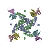
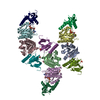
 Components
 Keywords
 Function and homology information
 Homo sapiens (human)
 X-RAY DIFFRACTION /
  Authors
 United States, 1items
  Citation
 Structure visualization
 Downloads & links
 Other downloads

 PDBj
PDBj Assembly


 Sample preparation
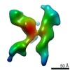
 Processing