[English] 日本語
Yorodumi
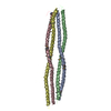
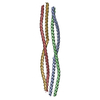
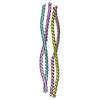
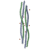
- PDB-6ec0: Crystal structure of the wild-type heterocomplex between coil 1B ... -

+
Open data


ID or keywords:

Loading...

-
Basic information

Entry
Database: PDB / ID: 6ec0
TitleCrystal structure of the wild-type heterocomplex between coil 1B domains of human intermediate filament proteins keratin 1 (KRT1) and keratin 10 (KRT10)
Components
  • Keratin 1
  • Keratin, type I cytoskeletal 10
KeywordsPROTEIN FIBRIL / keratin / intermediate filament / coiled-coil / skin
Function / homology
Function and homology information


positive regulation of epidermis development / structural constituent of skin epidermis / keratin filament / Keratinization / intermediate filament organization / Formation of the cornified envelope / peptide cross-linking / cornified envelope / protein heterotetramerization / intermediate filament ...positive regulation of epidermis development / structural constituent of skin epidermis / keratin filament / Keratinization / intermediate filament organization / Formation of the cornified envelope / peptide cross-linking / cornified envelope / protein heterotetramerization / intermediate filament / epidermis development / keratinocyte differentiation / epithelial cell differentiation / cytoskeleton / protein heterodimerization activity / cell surface / extracellular space / extracellular exosome / membrane / nucleus / plasma membrane / cytoplasm / cytosol
Similarity search - Function
Keratin type II cytoskeletal 1, tail / Keratin type II cytoskeletal 1 tail / Keratin, type I / Keratin, type II / Keratin type II head / Keratin type II head / : / Intermediate filament protein, conserved site / Intermediate filament protein / Intermediate filament (IF) rod domain signature. ...Keratin type II cytoskeletal 1, tail / Keratin type II cytoskeletal 1 tail / Keratin, type I / Keratin, type II / Keratin type II head / Keratin type II head / : / Intermediate filament protein, conserved site / Intermediate filament protein / Intermediate filament (IF) rod domain signature. / Intermediate filament, rod domain / Intermediate filament (IF) rod domain profile. / Intermediate filament protein
Similarity search - Domain/homology
: / : / NICKEL (II) ION / Cytokeratin-1 / Keratin, type I cytoskeletal 10
Similarity search - Component
Biological speciesHomo sapiens (human)
MethodX-RAY DIFFRACTION / SYNCHROTRON / MOLECULAR REPLACEMENT / molecular replacement / Resolution: 2.983 Å
AuthorsEldirany, S.A. / Lomakin, I.B. / Bunick, C.G.
Funding support United States, 2items
OrganizationGrant numberCountry
National Institutes of Health/National Institute of Arthritis and Musculoskeletal and Skin Diseases (NIH/NIAMS)K08-AR070290 United States
Other privateFoundation for Ichthyosis & Related Skin Types United States
CitationJournal: Embo J. / Year: 2019
Title: Human keratin 1/10-1B tetramer structures reveal a knob-pocket mechanism in intermediate filament assembly.
Authors: Eldirany, S.A. / Ho, M. / Hinbest, A.J. / Lomakin, I.B. / Bunick, C.G.
History
DepositionAug 7, 2018Deposition site: RCSB / Processing site: RCSB
Revision 1.0May 15, 2019Provider: repository / Type: Initial release
Revision 1.1Jun 12, 2019Group: Data collection / Database references / Category: citation / Item: _citation.journal_volume
Revision 1.2Dec 11, 2019Group: Author supporting evidence / Category: pdbx_audit_support / Item: _pdbx_audit_support.funding_organization
Revision 1.3Mar 13, 2024Group: Data collection / Database references / Derived calculations
Category: chem_comp_atom / chem_comp_bond ...chem_comp_atom / chem_comp_bond / database_2 / struct_conn
Item: _database_2.pdbx_DOI / _database_2.pdbx_database_accession ..._database_2.pdbx_DOI / _database_2.pdbx_database_accession / _struct_conn.pdbx_dist_value / _struct_conn.ptnr1_auth_asym_id / _struct_conn.ptnr1_auth_comp_id / _struct_conn.ptnr1_auth_seq_id / _struct_conn.ptnr1_label_asym_id / _struct_conn.ptnr1_label_atom_id / _struct_conn.ptnr1_label_comp_id / _struct_conn.ptnr1_label_seq_id / _struct_conn.ptnr1_symmetry / _struct_conn.ptnr2_auth_asym_id / _struct_conn.ptnr2_auth_comp_id / _struct_conn.ptnr2_auth_seq_id / _struct_conn.ptnr2_label_asym_id / _struct_conn.ptnr2_label_atom_id / _struct_conn.ptnr2_label_comp_id / _struct_conn.ptnr2_label_seq_id / _struct_conn.ptnr2_symmetry
Revision 1.4Apr 3, 2024Group: Refinement description / Category: pdbx_initial_refinement_model

-
Structure visualization

Structure viewerMolecule:
MolmilJmol/JSmol

Downloads & links

-
Assembly

Deposited unit
A: Keratin 1
B: Keratin, type I cytoskeletal 10
hetero molecules


Theoretical massNumber of molelcules
Total (without water)25,57110
Polymers24,8862
Non-polymers6858
Water63135
1
A: Keratin 1
B: Keratin, type I cytoskeletal 10
hetero molecules

A: Keratin 1
B: Keratin, type I cytoskeletal 10
hetero molecules


Theoretical massNumber of molelcules
Total (without water)51,14120
Polymers49,7714
Non-polymers1,37016
Water724
TypeNameSymmetry operationNumber
identity operation1_555x,y,z1
crystal symmetry operation6_555-x,-x+y,-z+1/31
Buried area16120 Å2
ΔGint-248 kcal/mol
Surface area27680 Å2
MethodPISA
Unit cell
Length a, b, c (Å)106.685, 106.685, 70.321
Angle α, β, γ (deg.)90.000, 90.000, 120.000
Int Tables number152
Space group name H-MP3121

-
Components

-
Protein , 2 types, 2 molecules AB

#1: Protein Keratin 1


Mass: 12764.193 Da / Num. of mol.: 1
Source method: isolated from a genetically manipulated source
Details: coil 1B domain of human keratin 1 (KRT1) / Source: (gene. exp.) Homo sapiens (human) / Gene: KRT1 / Plasmid: pET / Production host: Escherichia coli BL21(DE3) (bacteria) / Strain (production host): BL21(DE3) / References: UniProt: H6VRG1
#2: Protein Keratin, type I cytoskeletal 10 / Cytokeratin-10 / CK-10 / Keratin-10 / K10


Mass: 12121.534 Da / Num. of mol.: 1
Source method: isolated from a genetically manipulated source
Details: Gly-Ser at N-terminus following His-tag cleavageCoil 1B domain of human keratin 10 (KRT10)
Source: (gene. exp.) Homo sapiens (human) / Gene: KRT10, KPP / Plasmid: pET / Production host: Escherichia coli BL21(DE3) (bacteria) / Strain (production host): BL21(DE3) / References: UniProt: P13645

-
Non-polymers , 4 types, 43 molecules

#3: Chemical
ChemComp-CD / CADMIUM ION


Mass: 112.411 Da / Num. of mol.: 4 / Source method: obtained synthetically / Formula: Cd
#4: Chemical ChemComp-CO / COBALT (II) ION


Mass: 58.933 Da / Num. of mol.: 2 / Source method: obtained synthetically / Formula: Co
#5: Chemical ChemComp-NI / NICKEL (II) ION


Mass: 58.693 Da / Num. of mol.: 2 / Source method: obtained synthetically / Formula: Ni
#6: Water ChemComp-HOH / water


Mass: 18.015 Da / Num. of mol.: 35 / Source method: isolated from a natural source / Formula: H2O

-
Experimental details

-
Experiment

ExperimentMethod: X-RAY DIFFRACTION / Number of used crystals: 1

-
Sample preparation

CrystalDensity Matthews: 4.67 Å3/Da / Density % sol: 73.66 % / Description: thin, rod-shaped needles
Crystal growTemperature: 298 K / Method: vapor diffusion, sitting drop / pH: 7.5
Details: 11% PEG 3350, 0.1 M HEPES, 5 mM cobalt chloride, 5 mM cadmium chloride, 5 mM magnesium chloride, 5 mM nickel chloride

-
Data collection

DiffractionMean temperature: 100 K
Diffraction sourceSource: SYNCHROTRON / Site: APS / Beamline: 24-ID-C / Wavelength: 0.9795 Å
DetectorType: DECTRIS PILATUS 6M-F / Detector: PIXEL / Date: Jul 6, 2017
RadiationMonochromator: cryo-cooled double crystal Si(111) / Protocol: SINGLE WAVELENGTH / Monochromatic (M) / Laue (L): M / Scattering type: x-ray
Radiation wavelengthWavelength: 0.9795 Å / Relative weight: 1
ReflectionResolution: 2.983→50 Å / Num. obs: 8681 / % possible obs: 89.6 % / Redundancy: 8 % / Biso Wilson estimate: 91.24 Å2 / Rmerge(I) obs: 0.132 / Rpim(I) all: 0.047 / Rrim(I) all: 0.141 / Χ2: 1 / Net I/σ(I): 4.3
Reflection shell

Diffraction-ID: 1

Resolution (Å)Redundancy (%)Rmerge(I) obsNum. unique obsCC1/2Rpim(I) allRrim(I) allΧ2% possible all
2.983-3.0551.1852060.640.4771.2850.942.7
3.05-3.114.51.4622400.5310.6191.5970.92652.1
3.11-3.174.91.2332970.540.5421.3590.8862.5
3.17-3.235.11.2313270.6270.5281.3480.89269.6
3.23-3.35.31.3253830.6080.571.4540.81780.6
3.3-3.385.91.3244390.5480.551.4430.86588.5
3.38-3.466.30.8944400.8190.3660.9720.85695
3.46-3.566.70.6694740.9480.2650.7230.93398.1
3.56-3.666.60.5394670.9640.2160.5840.95599.4
3.66-3.787.30.4194890.9730.1610.4510.95299.6
3.78-3.918.90.3494810.9880.1230.3710.986100
3.91-4.079.60.3394730.9920.1150.3581.018100
4.07-4.26100.2494830.9940.0830.2621.065100
4.26-4.48100.1774850.9980.0590.1871.082100
4.48-4.769.90.154880.9990.0510.1591.142100
4.76-5.139.50.1074920.9980.0370.1141.133100
5.13-5.648.40.1444860.9960.0520.1531.072100
5.64-6.4610.20.1554980.9950.0510.1641.08899.8
6.46-8.139.80.0834970.9980.0280.0881.011100
8.13-508.80.055360.9990.0170.0530.822100

-
Phasing

PhasingMethod: molecular replacement

-
Processing

Software
NameVersionClassificationNB
PHENIXrefinement
HKL-2000data reduction
HKL-2000data scaling
MOLREPphasing
PDB_EXTRACT3.24data extraction
RefinementMethod to determine structure: MOLECULAR REPLACEMENT
Starting model: Phase problem initially solved using SAD on an isomorphous crystal whose data was collected at the cadmium edge (8500eV). This structure was used as starting model in MolRep to obtain ...Starting model: Phase problem initially solved using SAD on an isomorphous crystal whose data was collected at the cadmium edge (8500eV). This structure was used as starting model in MolRep to obtain the higher resolution KRT1/KRT10 1B structure.

Resolution: 2.983→46.196 Å / SU ML: 0.72 / Cross valid method: THROUGHOUT / σ(F): 1.34 / Phase error: 46.21
RfactorNum. reflection% reflectionSelection details
Rfree0.3001 398 4.73 %random selection
Rwork0.2814 ---
obs0.2822 8417 86.94 %-
Solvent computationShrinkage radii: 0.9 Å / VDW probe radii: 1.11 Å
Displacement parametersBiso max: 278.99 Å2 / Biso mean: 140.6592 Å2 / Biso min: 27.06 Å2
Refinement stepCycle: final / Resolution: 2.983→46.196 Å
ProteinNucleic acidLigandSolventTotal
Num. atoms1731 0 7 35 1773
Biso mean--163.86 118.11 -
Num. residues----210
Refine LS restraints
Refine-IDTypeDev idealNumber
X-RAY DIFFRACTIONf_bond_d0.0061746
X-RAY DIFFRACTIONf_angle_d0.8962347
X-RAY DIFFRACTIONf_chiral_restr0.036265
X-RAY DIFFRACTIONf_plane_restr0.004311
X-RAY DIFFRACTIONf_dihedral_angle_d18.411102
LS refinement shell

Refine-ID: X-RAY DIFFRACTION / Rfactor Rfree error: 0 / Total num. of bins used: 3

Resolution (Å)Rfactor RfreeNum. reflection RfreeRfactor RworkNum. reflection RworkNum. reflection all% reflection obs (%)
2.9832-3.41480.45821090.42421888199763
3.4148-4.30180.39091490.33942990313998
4.3018-46.20150.23971400.23843141328199
Refinement TLS params.

Method: refined / Refine-ID: X-RAY DIFFRACTION

IDL112)L122)L132)L222)L232)L332)S11 (Å °)S12 (Å °)S13 (Å °)S21 (Å °)S22 (Å °)S23 (Å °)S31 (Å °)S32 (Å °)S33 (Å °)T112)T122)T132)T222)T232)T332)Origin x (Å)Origin y (Å)Origin z (Å)
12.12250.5683-1.47730.5125-0.94171.85730.2290.1374-0.3055-0.25020.72140.01630.1102-0.28012.5960.6788-0.5859-0.30071.78410.38741.031256.7008-16.289865.3648
20.3879-0.0448-0.03920.00120.02620.1044-0.28310.26290.5774-0.3373-0.20960.3880.49350.76470.00521.174-0.2135-0.10791.70650.06790.757648.3587-21.020443.3649
30.4732-0.02150.09060.4801-0.67980.9367-0.58450.0940.1696-0.55990.24660.0698-0.76540.35410.00410.9995-0.1488-0.13260.9719-0.02690.806523.4407-28.92271.7834
40.24310.21380.46670.1810.40390.90220.69410.89610.73451.0076-0.7475-0.07351.74731.5939-0.01091.2604-0.1656-0.19711.6429-0.13240.8391-1.598-51.5841-39.0172
50.00020.0095-0.01270.0772-0.10850.14980.322-0.5429-0.19260.62910.7295-0.46350.5114-0.0603-0.00011.09310.0413-0.09551.34470.16561.3436-14.6133-59.1802-57.6145
60.17130.0479-0.01930.0616-0.10260.1925-0.86170.67261.99250.96830.79281.20520.6323-0.2245-0.00152.0388-0.44860.59571.4814-0.16351.802349.4395-9.532463.3701
70.0657-0.0944-0.07820.19410.19020.1009-0.72670.7582-0.06150.07640.55160.1831-0.05141.9533-0.047-0.0861-0.1942-0.41930.988-0.30480.337334.3662-24.294932.2508
80.3522-0.033-0.21970.422-0.14430.2383-0.36230.58970.40480.01610.8838-0.10730.76570.9315-0.00311.0274-0.0196-0.11361.8634-0.05040.679621.4526-39.4975-11.7265
90.6895-1.0836-0.27731.82470.65950.63070.34930.08490.1993-0.9679-1.5704-0.2710.698-0.4965-0.1291.8218-0.7486-0.11232.06570.16160.95696.8669-45.7223-41.3437
100.6689-0.84840.42861.0729-0.54420.27510.3791-0.5572-1.2896-0.559-0.04150.87670.6854-0.10640.12220.8767-0.1796-0.05181.1682-0.54680.7491-6.2505-49.1585-59.3196
Refinement TLS group
IDRefine-IDRefine TLS-IDSelection detailsAuth asym-IDAuth seq-ID
1X-RAY DIFFRACTION1(chain A and resid 225:232)A225 - 232
2X-RAY DIFFRACTION2(chain A and resid 233:255)A233 - 255
3X-RAY DIFFRACTION3(chain A and resid 256:299)A256 - 299
4X-RAY DIFFRACTION4(chain A and resid 300:325)A300 - 325
5X-RAY DIFFRACTION5(chain A and resid 326:331)A326 - 331
6X-RAY DIFFRACTION6(chain B and resid 195:201)B195 - 201
7X-RAY DIFFRACTION7(chain B and resid 202:243)B202 - 243
8X-RAY DIFFRACTION8(chain B and resid 244:266)B244 - 266
9X-RAY DIFFRACTION9(chain B and resid 267:287)B267 - 287
10X-RAY DIFFRACTION10(chain B and resid 288:296)B288 - 296

+
About Yorodumi

-
News

-
Feb 9, 2022. New format data for meta-information of EMDB entries

New format data for meta-information of EMDB entries

  • Version 3 of the EMDB header file is now the official format.
  • The previous official version 1.9 will be removed from the archive.

Related info.:EMDB header

External links:wwPDB to switch to version 3 of the EMDB data model

-
Aug 12, 2020. Covid-19 info

Covid-19 info

URL: https://pdbjlvh1.pdbj.org/emnavi/covid19.php

New page: Covid-19 featured information page in EM Navigator.

Related info.:Covid-19 info / Mar 5, 2020. Novel coronavirus structure data

+
Mar 5, 2020. Novel coronavirus structure data

Novel coronavirus structure data

Related info.:Yorodumi Speices / Aug 12, 2020. Covid-19 info

External links:COVID-19 featured content - PDBj / Molecule of the Month (242):Coronavirus Proteases

+
Jan 31, 2019. EMDB accession codes are about to change! (news from PDBe EMDB page)

EMDB accession codes are about to change! (news from PDBe EMDB page)

  • The allocation of 4 digits for EMDB accession codes will soon come to an end. Whilst these codes will remain in use, new EMDB accession codes will include an additional digit and will expand incrementally as the available range of codes is exhausted. The current 4-digit format prefixed with “EMD-” (i.e. EMD-XXXX) will advance to a 5-digit format (i.e. EMD-XXXXX), and so on. It is currently estimated that the 4-digit codes will be depleted around Spring 2019, at which point the 5-digit format will come into force.
  • The EM Navigator/Yorodumi systems omit the EMD- prefix.

Related info.:Q: What is EMD? / ID/Accession-code notation in Yorodumi/EM Navigator

External links:EMDB Accession Codes are Changing Soon! / Contact to PDBj

+
Jul 12, 2017. Major update of PDB

Major update of PDB

  • wwPDB released updated PDB data conforming to the new PDBx/mmCIF dictionary.
  • This is a major update changing the version number from 4 to 5, and with Remediation, in which all the entries are updated.
  • In this update, many items about electron microscopy experimental information are reorganized (e.g. em_software).
  • Now, EM Navigator and Yorodumi are based on the updated data.

External links:wwPDB Remediation / Enriched Model Files Conforming to OneDep Data Standards Now Available in the PDB FTP Archive

-
Yorodumi

Thousand views of thousand structures

  • Yorodumi is a browser for structure data from EMDB, PDB, SASBDB, etc.
  • This page is also the successor to EM Navigator detail page, and also detail information page/front-end page for Omokage search.
  • The word "yorodu" (or yorozu) is an old Japanese word meaning "ten thousand". "mi" (miru) is to see.

Related info.:EMDB / PDB / SASBDB / Comparison of 3 databanks / Yorodumi Search / Aug 31, 2016. New EM Navigator & Yorodumi / Yorodumi Papers / Jmol/JSmol / Function and homology information / Changes in new EM Navigator and Yorodumi

Read more