 Movie
Movie Controller

[English] 日本語
 Yorodumi
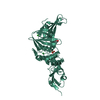
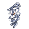
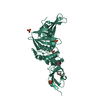
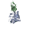
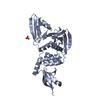
- PDB-5cag: Crystal structure of a putative adhesin (BACOVA_02677) from Bacte... -
+ Open data

- Basic information
| Entry | Database: PDB / ID: 5cag | ||||||
|---|---|---|---|---|---|---|---|
| Title | Crystal structure of a putative adhesin (BACOVA_02677) from Bacteroides ovatus ATCC 8483 at 3.00 A resolution (PSI Community Target, Nakayama) | ||||||
 Components Components | Uncharacterized protein | ||||||
 Keywords Keywords | CELL ADHESION / adhesin / Structural Genomics / Joint Center for Structural Genomics / JCSG / Protein Structure Initiative / PSI-BIOLOGY | ||||||
| Function / homology | Protein of unknown function DUF5119 / Domain of unknown function (DUF5119) / cell outer membrane / Prokaryotic membrane lipoprotein lipid attachment site profile. / Putative fimbrium anchoring subunit Fim4B Function and homology information Function and homology information | ||||||
| Biological species |  Bacteroides ovatus (bacteria) Bacteroides ovatus (bacteria) | ||||||
| Method |  X-RAY DIFFRACTION / SYNCHROTRON / MAD / Resolution: 3 Å X-RAY DIFFRACTION / SYNCHROTRON / MAD / Resolution: 3 Å | ||||||
 Authors Authors | Joint Center for Structural Genomics (JCSG) / Nakayama, K. | ||||||
 Citation Citation | Journal: Cell / Year: 2016 Title: A Distinct Type of Pilus from the Human Microbiome. Authors: Xu, Q. / Shoji, M. / Shibata, S. / Naito, M. / Sato, K. / Elsliger, M.A. / Grant, J.C. / Axelrod, H.L. / Chiu, H.J. / Farr, C.L. / Jaroszewski, L. / Knuth, M.W. / Deacon, A.M. / Godzik, A. / ...Authors: Xu, Q. / Shoji, M. / Shibata, S. / Naito, M. / Sato, K. / Elsliger, M.A. / Grant, J.C. / Axelrod, H.L. / Chiu, H.J. / Farr, C.L. / Jaroszewski, L. / Knuth, M.W. / Deacon, A.M. / Godzik, A. / Lesley, S.A. / Curtis, M.A. / Nakayama, K. / Wilson, I.A. | ||||||
| History |
|
- Structure visualization
| Structure viewer | Molecule: MolmilJmol/JSmol |
|---|
- Downloads & links
-Download
| PDBx/mmCIF format | 5cag.cif.gz | 130.4 KB | Display | PDBx/mmCIF format |
|---|---|---|---|---|
| PDB format | pdb5cag.ent.gz | 102.2 KB | Display | PDB format |
| PDBx/mmJSON format | 5cag.json.gz | Tree view | PDBx/mmJSON format | |
| Others |  Other downloads Other downloads |
-Validation report
| Arichive directory | https://data.pdbj.org/pub/pdb/validation_reports/ca/5cagftp://data.pdbj.org/pub/pdb/validation_reports/ca/5cag | HTTPS FTP |
|---|
-Related structure data
| Related structure data |  3liuC  3payC  3r4rC  3sy6C 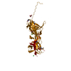 3t2lC 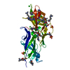 3ufiC  3up6C 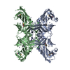 4dguC  4epsC 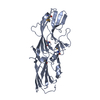 4gpvC 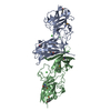 4h40C  4jg5C  4jrfC  4k4kC  4q98C 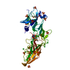 4qb7C  4qdgC  4rdbC C: citing same article ( |
|---|---|
| Similar structure data | |
| Other databases |


-Links
 PDBj
PDBj


- Assembly
| Deposited unit | 
| ||||||||
|---|---|---|---|---|---|---|---|---|---|
| 1 |
| ||||||||
| 2 | 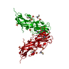
| ||||||||
| Unit cell |
|
-Components
| #1: Protein | Mass: 35280.500 Da / Num. of mol.: 1 Source method: isolated from a genetically manipulated source Source: (gene. exp.) Bacteroides ovatus (bacteria) / Strain: ATCC 8483 / DSM 1896 / JCM 5824 / NCTC 11153 / Gene: BACOVA_02677 / Plasmid: SpeedET / Production host: | ||
|---|---|---|---|
| #2: Chemical | ChemComp-SO4 /   Mass: 96.063 Da / Num. of mol.: 9 / Source method: obtained synthetically / Formula: SO4 Mass: 96.063 Da / Num. of mol.: 9 / Source method: obtained synthetically / Formula: SO4Has protein modification | Y | |
-Experimental details
-Experiment
| Experiment | Method: X-RAY DIFFRACTION / Number of used crystals: 1 |
|---|
- Sample preparation
| Crystal | Density Matthews: 4.3 Å3/Da / Density % sol: 71.38 % |
|---|---|
| Crystal grow | Temperature: 277 K / Method: vapor diffusion, sitting drop / pH: 10.5 Details: 0.2000M lithium sulfate, 2.0000M ammonium sulfate, 0.1M CAPS pH 10.5 |
-Data collection
| Diffraction | Mean temperature: 100 K | ||||||||||||||||||||||||||||||||||||||||||||||||||||||||||||||||||||||||||||||||||||||||||||||||||||||||||||||||||||||||||||||||||||
|---|---|---|---|---|---|---|---|---|---|---|---|---|---|---|---|---|---|---|---|---|---|---|---|---|---|---|---|---|---|---|---|---|---|---|---|---|---|---|---|---|---|---|---|---|---|---|---|---|---|---|---|---|---|---|---|---|---|---|---|---|---|---|---|---|---|---|---|---|---|---|---|---|---|---|---|---|---|---|---|---|---|---|---|---|---|---|---|---|---|---|---|---|---|---|---|---|---|---|---|---|---|---|---|---|---|---|---|---|---|---|---|---|---|---|---|---|---|---|---|---|---|---|---|---|---|---|---|---|---|---|---|---|---|
| Diffraction source | Source: SYNCHROTRON / Site: SSRL  / Beamline: BL12-2 / Wavelength: 0.9116,0.9794,0.9792 / Beamline: BL12-2 / Wavelength: 0.9116,0.9794,0.9792 | ||||||||||||||||||||||||||||||||||||||||||||||||||||||||||||||||||||||||||||||||||||||||||||||||||||||||||||||||||||||||||||||||||||
| Detector | Type: DECTRIS PILATUS 6M / Detector: PIXEL / Date: Jul 28, 2014 Details: Rhodium-coated vertical and horizontal focusing mirrors; liquid-nitrogen cooled double crystal Si(111) monochromator | ||||||||||||||||||||||||||||||||||||||||||||||||||||||||||||||||||||||||||||||||||||||||||||||||||||||||||||||||||||||||||||||||||||
| Radiation | Monochromator: double crystal Si(111) / Protocol: MAD / Monochromatic (M) / Laue (L): M / Scattering type: x-ray | ||||||||||||||||||||||||||||||||||||||||||||||||||||||||||||||||||||||||||||||||||||||||||||||||||||||||||||||||||||||||||||||||||||
| Radiation wavelength |
| ||||||||||||||||||||||||||||||||||||||||||||||||||||||||||||||||||||||||||||||||||||||||||||||||||||||||||||||||||||||||||||||||||||
| Reflection | Resolution: 3→49.195 Å / Num. obs: 13098 / % possible obs: 99.8 % / Observed criterion σ(I): -3 / Redundancy: 9.491 % / Biso Wilson estimate: 81.271 Å2 / Rmerge F obs: 0.999 / Rmerge(I) obs: 0.092 / Rrim(I) all: 0.098 / Net I/σ(I): 18.18 / Num. measured all: 125002 | ||||||||||||||||||||||||||||||||||||||||||||||||||||||||||||||||||||||||||||||||||||||||||||||||||||||||||||||||||||||||||||||||||||
| Reflection shell |
|
-Phasing
| Phasing | Method: MAD |
|---|
- Processing
| Software |
| ||||||||||||||||||||||||||||||||||||||||||||||||||||||||||||||||||||||||||||||||||||||||||||||||||||||||||||
|---|---|---|---|---|---|---|---|---|---|---|---|---|---|---|---|---|---|---|---|---|---|---|---|---|---|---|---|---|---|---|---|---|---|---|---|---|---|---|---|---|---|---|---|---|---|---|---|---|---|---|---|---|---|---|---|---|---|---|---|---|---|---|---|---|---|---|---|---|---|---|---|---|---|---|---|---|---|---|---|---|---|---|---|---|---|---|---|---|---|---|---|---|---|---|---|---|---|---|---|---|---|---|---|---|---|---|---|---|---|
| Refinement | Method to determine structure: MAD / Resolution: 3→49.195 Å / Cor.coef. Fo:Fc: 0.9216 / Cor.coef. Fo:Fc free: 0.9328 / Occupancy max: 1 / Occupancy min: 0.5 / Cross valid method: THROUGHOUT / σ(F): 0 Details: 1. A MET-INHIBITION PROTOCOL WAS USED FOR SELENOMETHIONINE INCORPORATION DURING PROTEIN EXPRESSION. THE OCCUPANCY OF THE SE ATOMS IN THE MSE RESIDUES WAS REDUCED TO 0.75 FOR THE REDUCED ...Details: 1. A MET-INHIBITION PROTOCOL WAS USED FOR SELENOMETHIONINE INCORPORATION DURING PROTEIN EXPRESSION. THE OCCUPANCY OF THE SE ATOMS IN THE MSE RESIDUES WAS REDUCED TO 0.75 FOR THE REDUCED SCATTERING POWER DUE TO PARTIAL S-MET INCORPORATION. 2. THE MAD PHASES WERE USED AS RESTRAINTS DURING REFINEMENT. 3. HYDROGENS HAVE BEEN ADDED IN THE RIDING POSITIONS. 4. ATOM RECORDS CONTAIN SUM OF TLS AND RESIDUAL B FACTORS. ANISOU RECORDS CONTAIN SUM OF TLS AND RESIDUAL U FACTORS. 5. THE SEQUENCE IS NUMBERED ACCORDING PATRIC DB ENTRY VBIBacOva42630_2252 WHICH CONTAINS ADDITIONAL N-TERM SIGNAL SEQUENCE (NOT PRESENT IN UNIPROT ENTRY VBIBacOva42630_2252).
| ||||||||||||||||||||||||||||||||||||||||||||||||||||||||||||||||||||||||||||||||||||||||||||||||||||||||||||
| Displacement parameters | Biso max: 225.89 Å2 / Biso mean: 107.9401 Å2 / Biso min: 59.6 Å2
| ||||||||||||||||||||||||||||||||||||||||||||||||||||||||||||||||||||||||||||||||||||||||||||||||||||||||||||
| Refine analyze | Luzzati coordinate error obs: 0.564 Å | ||||||||||||||||||||||||||||||||||||||||||||||||||||||||||||||||||||||||||||||||||||||||||||||||||||||||||||
| Refinement step | Cycle: LAST / Resolution: 3→49.195 Å
| ||||||||||||||||||||||||||||||||||||||||||||||||||||||||||||||||||||||||||||||||||||||||||||||||||||||||||||
| Refine LS restraints |
| ||||||||||||||||||||||||||||||||||||||||||||||||||||||||||||||||||||||||||||||||||||||||||||||||||||||||||||
| LS refinement shell | Resolution: 3→3.24 Å / Total num. of bins used: 7
| ||||||||||||||||||||||||||||||||||||||||||||||||||||||||||||||||||||||||||||||||||||||||||||||||||||||||||||
| Refinement TLS params. | Method: refined / Origin x: 25.9473 Å / Origin y: 60.0926 Å / Origin z: 117.583 Å
| ||||||||||||||||||||||||||||||||||||||||||||||||||||||||||||||||||||||||||||||||||||||||||||||||||||||||||||
| Refinement TLS group | Selection details: {A|0 - 306} |
