 Movie
Movie Controller

[English] 日本語
 Yorodumi
- PDB-4eps: Crystal structure of a fimbrial protein (BACOVA_04982) from Bacte... -
+ Open data

- Basic information
| Entry | Database: PDB / ID: 4eps | ||||||
|---|---|---|---|---|---|---|---|
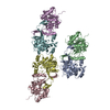
| Title | Crystal structure of a fimbrial protein (BACOVA_04982) from Bacteroides ovatus ATCC 8483 at 1.85 A resolution | ||||||
 Components Components | hypothetical protein | ||||||
 Keywords Keywords | Structural Genomics / Unknown Function / HYPOTHETICAL PROTEIN (DUF3988) / JOINT CENTER FOR STRUCTURAL GENOMICS / JCSG / PROTEIN STRUCTURE INITIATIVE / PSI-BIOLOGY | ||||||
| Function / homology |  Function and homology information Function and homology information | ||||||
| Biological species |  Bacteroides ovatus (bacteria) Bacteroides ovatus (bacteria) | ||||||
| Method |  X-RAY DIFFRACTION / SYNCHROTRON / SAD / Resolution: 1.85 Å X-RAY DIFFRACTION / SYNCHROTRON / SAD / Resolution: 1.85 Å | ||||||
 Authors Authors | Joint Center for Structural Genomics (JCSG) | ||||||
 Citation Citation | Journal: Cell / Year: 2016 Title: A Distinct Type of Pilus from the Human Microbiome. Authors: Xu, Q. / Shoji, M. / Shibata, S. / Naito, M. / Sato, K. / Elsliger, M.A. / Grant, J.C. / Axelrod, H.L. / Chiu, H.J. / Farr, C.L. / Jaroszewski, L. / Knuth, M.W. / Deacon, A.M. / Godzik, A. / ...Authors: Xu, Q. / Shoji, M. / Shibata, S. / Naito, M. / Sato, K. / Elsliger, M.A. / Grant, J.C. / Axelrod, H.L. / Chiu, H.J. / Farr, C.L. / Jaroszewski, L. / Knuth, M.W. / Deacon, A.M. / Godzik, A. / Lesley, S.A. / Curtis, M.A. / Nakayama, K. / Wilson, I.A. | ||||||
| History |
|
- Structure visualization
| Structure viewer | Molecule: MolmilJmol/JSmol |
|---|
- Downloads & links
-Download
| PDBx/mmCIF format | 4eps.cif.gz | 239.5 KB | Display | PDBx/mmCIF format |
|---|---|---|---|---|
| PDB format | pdb4eps.ent.gz | 190 KB | Display | PDB format |
| PDBx/mmJSON format | 4eps.json.gz | Tree view | PDBx/mmJSON format | |
| Others |  Other downloads Other downloads |
-Validation report
| Arichive directory | https://data.pdbj.org/pub/pdb/validation_reports/ep/4epsftp://data.pdbj.org/pub/pdb/validation_reports/ep/4eps | HTTPS FTP |
|---|
-Related structure data
| Related structure data |  3liuC  3payC  3r4rC  3sy6C  3t2lC  3ufiC  3up6C  4dguC  4gpvC  4h40C  4jg5C  4jrfC  4k4kC  4q98C  4qb7C  4qdgC  4rdbC  5cagC C: citing same article ( |
|---|---|
| Similar structure data | |
| Other databases |


-Links
 PDBj
PDBj


- Assembly
| Deposited unit | 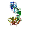
| ||||||||
|---|---|---|---|---|---|---|---|---|---|
| 1 | 
| ||||||||
| Unit cell |
|
-Components
| #1: Protein | Mass: 57519.613 Da / Num. of mol.: 1 Source method: isolated from a genetically manipulated source Source: (gene. exp.) Bacteroides ovatus (bacteria) / Strain: ATCC 8483 / Gene: BACOVA_04982 / Plasmid: SpeedET / Production host: | ||||||||||
|---|---|---|---|---|---|---|---|---|---|---|---|
| #2: Chemical | ChemComp-CL /   Mass: 35.453 Da / Num. of mol.: 4 / Source method: obtained synthetically / Formula: Cl Mass: 35.453 Da / Num. of mol.: 4 / Source method: obtained synthetically / Formula: Cl#3: Chemical |   Mass: 96.063 Da / Num. of mol.: 3 / Source method: obtained synthetically / Formula: SO4 Mass: 96.063 Da / Num. of mol.: 3 / Source method: obtained synthetically / Formula: SO4#4: Chemical | ChemComp-GOL /   Mass: 92.094 Da / Num. of mol.: 22 / Source method: obtained synthetically / Formula: C3H8O3 Mass: 92.094 Da / Num. of mol.: 22 / Source method: obtained synthetically / Formula: C3H8O3#5: Water | ChemComp-HOH / |  Mass: 18.015 Da / Num. of mol.: 656 / Source method: isolated from a natural source / Formula: H2O Mass: 18.015 Da / Num. of mol.: 656 / Source method: isolated from a natural source / Formula: H2OHas protein modification | Y | Sequence details | THE CONSTRUCT (RESIDUES 26-534) WAS EXPRESSED WITH A PURIFICATION TAG MGSDKIHHHHHHENLYFQG. THE TAG ...THE CONSTRUCT (RESIDUES 26-534) WAS EXPRESSED WITH A PURIFICATI | |
-Experimental details
-Experiment
| Experiment | Method: X-RAY DIFFRACTION / Number of used crystals: 1 |
|---|
- Sample preparation
| Crystal | Density Matthews: 3.31 Å3/Da / Density % sol: 62.82 % |
|---|---|
| Crystal grow | Temperature: 293 K / Method: vapor diffusion, sitting drop / pH: 6 Details: 1.6M ammonium sulfate, 0.1M MES pH 6.0, NANODROP, VAPOR DIFFUSION, SITTING DROP, temperature 293K |
-Data collection
| Diffraction | Mean temperature: 100 K | ||||||||||||||||||||||||||||||||||||||||||||||||||||||||||||||||||||||||||||||||||||||||||||||||||||||||||||||||||||||||||||||||||||||||||||||||||||||||||||||||||||||||
|---|---|---|---|---|---|---|---|---|---|---|---|---|---|---|---|---|---|---|---|---|---|---|---|---|---|---|---|---|---|---|---|---|---|---|---|---|---|---|---|---|---|---|---|---|---|---|---|---|---|---|---|---|---|---|---|---|---|---|---|---|---|---|---|---|---|---|---|---|---|---|---|---|---|---|---|---|---|---|---|---|---|---|---|---|---|---|---|---|---|---|---|---|---|---|---|---|---|---|---|---|---|---|---|---|---|---|---|---|---|---|---|---|---|---|---|---|---|---|---|---|---|---|---|---|---|---|---|---|---|---|---|---|---|---|---|---|---|---|---|---|---|---|---|---|---|---|---|---|---|---|---|---|---|---|---|---|---|---|---|---|---|---|---|---|---|---|---|---|---|
| Diffraction source | Source: SYNCHROTRON / Site: SSRL  / Beamline: BL12-2 / Wavelength: 0.9794 / Beamline: BL12-2 / Wavelength: 0.9794 | ||||||||||||||||||||||||||||||||||||||||||||||||||||||||||||||||||||||||||||||||||||||||||||||||||||||||||||||||||||||||||||||||||||||||||||||||||||||||||||||||||||||||
| Detector | Type: DECTRIS PILATUS 6M / Detector: PIXEL / Date: Mar 10, 2012 Details: Rhodium-coated vertical and horizontal focusing mirrors; liquid-nitrogen cooled double crystal Si(111) monochromator | ||||||||||||||||||||||||||||||||||||||||||||||||||||||||||||||||||||||||||||||||||||||||||||||||||||||||||||||||||||||||||||||||||||||||||||||||||||||||||||||||||||||||
| Radiation | Monochromator: double crystal Si(111) / Protocol: SINGLE WAVELENGTH / Monochromatic (M) / Laue (L): M / Scattering type: x-ray | ||||||||||||||||||||||||||||||||||||||||||||||||||||||||||||||||||||||||||||||||||||||||||||||||||||||||||||||||||||||||||||||||||||||||||||||||||||||||||||||||||||||||
| Radiation wavelength | Wavelength: 0.9794 Å / Relative weight: 1 | ||||||||||||||||||||||||||||||||||||||||||||||||||||||||||||||||||||||||||||||||||||||||||||||||||||||||||||||||||||||||||||||||||||||||||||||||||||||||||||||||||||||||
| Reflection | Resolution: 1.85→29.056 Å / Num. all: 67064 / Num. obs: 67064 / % possible obs: 99.9 % / Redundancy: 8.7 % / Rsym value: 0.114 / Net I/σ(I): 10.5 | ||||||||||||||||||||||||||||||||||||||||||||||||||||||||||||||||||||||||||||||||||||||||||||||||||||||||||||||||||||||||||||||||||||||||||||||||||||||||||||||||||||||||
| Reflection shell | Diffraction-ID: 1
|
-Phasing
| Phasing | Method: SAD |
|---|
- Processing
| Software |
| ||||||||||||||||||||||||||||||||||||||||||||||||||||||||||||||||||||||||||||||||||||||||||||||||||||||||||||
|---|---|---|---|---|---|---|---|---|---|---|---|---|---|---|---|---|---|---|---|---|---|---|---|---|---|---|---|---|---|---|---|---|---|---|---|---|---|---|---|---|---|---|---|---|---|---|---|---|---|---|---|---|---|---|---|---|---|---|---|---|---|---|---|---|---|---|---|---|---|---|---|---|---|---|---|---|---|---|---|---|---|---|---|---|---|---|---|---|---|---|---|---|---|---|---|---|---|---|---|---|---|---|---|---|---|---|---|---|---|
| Refinement | Method to determine structure: SAD / Resolution: 1.85→29.056 Å / Cor.coef. Fo:Fc: 0.9617 / Cor.coef. Fo:Fc free: 0.9584 / Occupancy max: 1 / Occupancy min: 0.3 / Cross valid method: THROUGHOUT / σ(F): 0 Details: 1. A MET-INHIBITION PROTOCOL WAS USED FOR SELENOMETHIONINE INCORPORATION DURING PROTEIN EXPRESSION. THE OCCUPANCY OF THE SE ATOMS IN THE MSE RESIDUES WAS REDUCED TO 0.75 FOR THE REDUCED ...Details: 1. A MET-INHIBITION PROTOCOL WAS USED FOR SELENOMETHIONINE INCORPORATION DURING PROTEIN EXPRESSION. THE OCCUPANCY OF THE SE ATOMS IN THE MSE RESIDUES WAS REDUCED TO 0.75 FOR THE REDUCED SCATTERING POWER DUE TO PARTIAL S-MET INCORPORATION. 2. ATOM RECORD CONTAINS SUM OF TLS AND RESIDUAL B FACTORS. ANISOU RECORD CONTAINS SUM OF TLS AND RESIDUAL U FACTORS. 3. THE MAD PHASES WERE USED AS RESTRAINTS DURING REFINEMENT. 4. SULFATE, CHLORIDE, GLYCEROL MODELED WERE PRESENT IN THE CRYSTALLIZATION/CRYO CONDITIONS.
| ||||||||||||||||||||||||||||||||||||||||||||||||||||||||||||||||||||||||||||||||||||||||||||||||||||||||||||
| Displacement parameters | Biso max: 114.12 Å2 / Biso mean: 37.2853 Å2 / Biso min: 15.58 Å2
| ||||||||||||||||||||||||||||||||||||||||||||||||||||||||||||||||||||||||||||||||||||||||||||||||||||||||||||
| Refine analyze | Luzzati coordinate error obs: 0.197 Å | ||||||||||||||||||||||||||||||||||||||||||||||||||||||||||||||||||||||||||||||||||||||||||||||||||||||||||||
| Refinement step | Cycle: LAST / Resolution: 1.85→29.056 Å
| ||||||||||||||||||||||||||||||||||||||||||||||||||||||||||||||||||||||||||||||||||||||||||||||||||||||||||||
| Refine LS restraints |
| ||||||||||||||||||||||||||||||||||||||||||||||||||||||||||||||||||||||||||||||||||||||||||||||||||||||||||||
| LS refinement shell | Resolution: 1.85→1.9 Å / Total num. of bins used: 20
| ||||||||||||||||||||||||||||||||||||||||||||||||||||||||||||||||||||||||||||||||||||||||||||||||||||||||||||
| Refinement TLS params. | Method: refined / Origin x: 46.4263 Å / Origin y: 34.1448 Å / Origin z: 92.1478 Å
| ||||||||||||||||||||||||||||||||||||||||||||||||||||||||||||||||||||||||||||||||||||||||||||||||||||||||||||
| Refinement TLS group | Selection details: { A|32 - 534 } |
