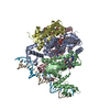
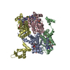
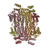
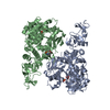
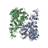
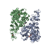
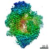
Entry Database : PDB / ID : 5c8eTitle Crystal structure of Thermus thermophilus CarH bound to adenosylcobalamin and a 26-bp DNA segment 26-mer DNA segment containing the CarH operator sequence (antisense strand) 26-mer DNA segment containing the CarH operator sequence (sense strand) Light-dependent transcriptional regulator CarH Keywords / / / / / Function / homology Function Domain/homology Component
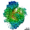
/ / / / / / / / / / / / / / / / / / / / / / / / / / / / / / / / / / Biological species Thermus thermophilus (bacteria)Thermus thermophilus HB27 (bacteria)Method / / / Resolution : 3.89 Å Authors Jost, M. / Drennan, C.L. Funding support Organization Grant number Country National Institutes of Health/National Institute of General Medical Sciences (NIH/NIGMS) GM069857 Howard Hughes Medical Institute (HHMI)
Journal : Nature / Year : 2015Title : Structural basis for gene regulation by a B12-dependent photoreceptor.Authors : Jost, M. / Fernandez-Zapata, J. / Polanco, M.C. / Ortiz-Guerrero, J.M. / Chen, P.Y. / Kang, G. / Padmanabhan, S. / Elias-Arnanz, M. / Drennan, C.L. History Deposition Jun 25, 2015 Deposition site / Processing site Revision 1.0 Sep 30, 2015 Provider / Type Revision 1.1 Oct 14, 2015 Group Revision 1.2 Nov 4, 2015 Group Revision 1.3 Sep 20, 2017 Group / Database references / Derived calculationsCategory / pdbx_audit_support / pdbx_struct_oper_listItem / _pdbx_audit_support.funding_organization / _pdbx_struct_oper_list.symmetry_operationRevision 1.4 Nov 20, 2019 Group / Category / Item Revision 1.5 Sep 27, 2023 Group Data collection / Database references ... Data collection / Database references / Derived calculations / Refinement description Category chem_comp_atom / chem_comp_bond ... chem_comp_atom / chem_comp_bond / database_2 / pdbx_initial_refinement_model / struct_conn Item _database_2.pdbx_DOI / _database_2.pdbx_database_accession ... _database_2.pdbx_DOI / _database_2.pdbx_database_accession / _struct_conn.pdbx_dist_value / _struct_conn.ptnr1_auth_asym_id / _struct_conn.ptnr1_auth_comp_id / _struct_conn.ptnr1_auth_seq_id / _struct_conn.ptnr1_label_asym_id / _struct_conn.ptnr1_label_atom_id / _struct_conn.ptnr1_label_comp_id / _struct_conn.ptnr1_label_seq_id / _struct_conn.ptnr2_auth_asym_id / _struct_conn.ptnr2_auth_comp_id / _struct_conn.ptnr2_auth_seq_id / _struct_conn.ptnr2_label_asym_id / _struct_conn.ptnr2_label_atom_id / _struct_conn.ptnr2_label_comp_id
Show all Show less
 Movie
Movie Controller

 Yorodumi
 Open data
 Basic information
 Components
 Keywords
 Function and homology information
 Thermus thermophilus (bacteria)
 X-RAY DIFFRACTION /
  Authors
 United States, 2items
  Citation
 Structure visualization
 Downloads & links
 Other downloads


 PDBj
PDBj


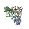
 Assembly