 Movie
Movie Controller

[English] 日本語
 Yorodumi
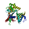
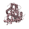
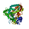
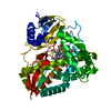
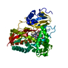
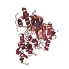
- PDB-5c7t: Crystal Structure of the Bdellovibrio bacteriovorus Nucleoside Di... -
+ Open data

- Basic information
| Entry | Database: PDB / ID: 5c7t | ||||||
|---|---|---|---|---|---|---|---|
| Title | Crystal Structure of the Bdellovibrio bacteriovorus Nucleoside Diphosphate Sugar Hydrolase in complex with ADP-ribose | ||||||
 Components Components | NudF protein | ||||||
 Keywords Keywords | HYDROLASE / Nudix | ||||||
| Function / homology |  Function and homology information Function and homology informationnucleoside phosphate metabolic process / ribose phosphate metabolic process / hydrolase activity / cytosol Similarity search - Function | ||||||
| Biological species |  Bdellovibrio bacteriovorus (bacteria) Bdellovibrio bacteriovorus (bacteria) | ||||||
| Method |  X-RAY DIFFRACTION / FOURIER SYNTHESIS / Resolution: 2.06 Å X-RAY DIFFRACTION / FOURIER SYNTHESIS / Resolution: 2.06 Å | ||||||
 Authors Authors | Gabelli, S.B. / de la Pena, A.H. / Suarez, A. / Amzel, L.M. | ||||||
 Citation Citation | Journal: Plos One / Year: 2015 Title: Structural and Enzymatic Characterization of a Nucleoside Diphosphate Sugar Hydrolase from Bdellovibrio bacteriovorus. Authors: de la Pena, A.H. / Suarez, A. / Duong-Ly, K.C. / Schoeffield, A.J. / Pizarro-Dupuy, M.A. / Zarr, M. / Pineiro, S.A. / Amzel, L.M. / Gabelli, S.B. | ||||||
| History |
|
- Structure visualization
| Structure viewer | Molecule: MolmilJmol/JSmol |
|---|
- Downloads & links
-Download
| PDBx/mmCIF format | 5c7t.cif.gz | 99.4 KB | Display | PDBx/mmCIF format |
|---|---|---|---|---|
| PDB format | pdb5c7t.ent.gz | 74.5 KB | Display | PDB format |
| PDBx/mmJSON format | 5c7t.json.gz | Tree view | PDBx/mmJSON format | |
| Others |  Other downloads Other downloads |
-Validation report
| Arichive directory | https://data.pdbj.org/pub/pdb/validation_reports/c7/5c7tftp://data.pdbj.org/pub/pdb/validation_reports/c7/5c7t | HTTPS FTP |
|---|
-Related structure data
| Related structure data | 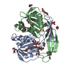 5c7qSC 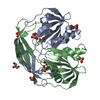 5c8lC S: Starting model for refinement C: citing same article ( |
|---|---|
| Similar structure data |


-Links
 PDBj
PDBj


- Assembly
| Deposited unit | 
| ||||||||
|---|---|---|---|---|---|---|---|---|---|
| 1 |
| ||||||||
| Unit cell |
|
-Components
-Protein , 1 types, 2 molecules AB
| #1: Protein | Mass: 21290.469 Da / Num. of mol.: 2 / Mutation: E140Q Source method: isolated from a genetically manipulated source Source: (gene. exp.) Bdellovibrio bacteriovorus (bacteria) / Strain: ATCC 15356 / DSM 50701 / NCIB 9529 / HD100 / Gene: nudF, Bd3179 / Plasmid: pET24Production host: References: UniProt: Q6MIH8, ADP-ribose diphosphatase |
|---|
-Non-polymers , 7 types, 230 molecules 


| #2: Chemical |  Mass: 559.316 Da / Num. of mol.: 2 / Source method: obtained synthetically / Formula: C15H23N5O14P2 Mass: 559.316 Da / Num. of mol.: 2 / Source method: obtained synthetically / Formula: C15H23N5O14P2#3: Chemical | ChemComp-SO4 /  Mass: 96.063 Da / Num. of mol.: 6 / Source method: obtained synthetically / Formula: SO4 Mass: 96.063 Da / Num. of mol.: 6 / Source method: obtained synthetically / Formula: SO4#4: Chemical | ChemComp-GOL /  Mass: 92.094 Da / Num. of mol.: 7 / Source method: obtained synthetically / Formula: C3H8O3 Mass: 92.094 Da / Num. of mol.: 7 / Source method: obtained synthetically / Formula: C3H8O3#5: Chemical | ChemComp-PG0 / |  Mass: 120.147 Da / Num. of mol.: 1 / Source method: obtained synthetically / Formula: C5H12O3 / Comment: precipitant*YM Mass: 120.147 Da / Num. of mol.: 1 / Source method: obtained synthetically / Formula: C5H12O3 / Comment: precipitant*YM#6: Chemical | ChemComp-PEG / |  Mass: 106.120 Da / Num. of mol.: 1 / Source method: obtained synthetically / Formula: C4H10O3 Mass: 106.120 Da / Num. of mol.: 1 / Source method: obtained synthetically / Formula: C4H10O3#7: Chemical | ChemComp-PGE / |  Mass: 150.173 Da / Num. of mol.: 1 / Source method: obtained synthetically / Formula: C6H14O4 Mass: 150.173 Da / Num. of mol.: 1 / Source method: obtained synthetically / Formula: C6H14O4#8: Water | ChemComp-HOH / | Mass: 18.015 Da / Num. of mol.: 212 / Source method: isolated from a natural source / Formula: H2O |
|---|
-Experimental details
-Experiment
| Experiment | Method: X-RAY DIFFRACTION / Number of used crystals: 1 |
|---|
- Sample preparation
| Crystal | Density Matthews: 2.37 Å3/Da / Density % sol: 48.08 % |
|---|---|
| Crystal grow | Temperature: 293 K / Method: vapor diffusion, hanging drop / pH: 7 Details: 1.75-2.0 M ammonium sulfate, 0.1 M HEPES pH 7.0, and 0-0.5% PEG 8000 |
-Data collection
| Diffraction | Mean temperature: 93 K | ||||||||||||||||||||||||||||||||||||||||||||||||||||||||||||||||||
|---|---|---|---|---|---|---|---|---|---|---|---|---|---|---|---|---|---|---|---|---|---|---|---|---|---|---|---|---|---|---|---|---|---|---|---|---|---|---|---|---|---|---|---|---|---|---|---|---|---|---|---|---|---|---|---|---|---|---|---|---|---|---|---|---|---|---|---|
| Diffraction source | Source: ROTATING ANODE / Type: RIGAKU FR-E+ DW / Wavelength: 1.541 Å | ||||||||||||||||||||||||||||||||||||||||||||||||||||||||||||||||||
| Detector | Type: RIGAKU SATURN 944+ / Detector: CCD / Date: Jun 24, 2010 | ||||||||||||||||||||||||||||||||||||||||||||||||||||||||||||||||||
| Radiation | Protocol: SINGLE WAVELENGTH / Monochromatic (M) / Laue (L): M / Scattering type: x-ray | ||||||||||||||||||||||||||||||||||||||||||||||||||||||||||||||||||
| Radiation wavelength | Wavelength: 1.541 Å / Relative weight: 1 | ||||||||||||||||||||||||||||||||||||||||||||||||||||||||||||||||||
| Reflection | Resolution: 2.06→50 Å / Num. obs: 24431 / % possible obs: 95 % / Redundancy: 5.4 % / Rmerge(I) obs: 0.095 / Χ2: 1.874 / Net I/av σ(I): 17.608 / Net I/σ(I): 11.9 / Num. measured all: 130858 | ||||||||||||||||||||||||||||||||||||||||||||||||||||||||||||||||||
| Reflection shell | Diffraction-ID: 1 / Rejects: _
|
- Processing
| Software |
| |||||||||||||||||||||||||||||||||||||||||||||||||||||||||||||||||||||||||||
|---|---|---|---|---|---|---|---|---|---|---|---|---|---|---|---|---|---|---|---|---|---|---|---|---|---|---|---|---|---|---|---|---|---|---|---|---|---|---|---|---|---|---|---|---|---|---|---|---|---|---|---|---|---|---|---|---|---|---|---|---|---|---|---|---|---|---|---|---|---|---|---|---|---|---|---|---|
| Refinement | Method to determine structure: FOURIER SYNTHESIS Starting model: 5c7Q Resolution: 2.06→26.8 Å / Cor.coef. Fo:Fc: 0.953 / Cor.coef. Fo:Fc free: 0.925 / WRfactor Rfree: 0.2342 / WRfactor Rwork: 0.1673 / FOM work R set: 0.8043 / SU B: 5.494 / SU ML: 0.143 / SU R Cruickshank DPI: 0.2291 / SU Rfree: 0.1999 / Cross valid method: THROUGHOUT / σ(F): 0 / ESU R: 0.229 / ESU R Free: 0.2 / Stereochemistry target values: MAXIMUM LIKELIHOOD Details: HYDROGENS HAVE BEEN ADDED IN THE RIDING POSITIONS U VALUES : REFINED INDIVIDUALLY
| |||||||||||||||||||||||||||||||||||||||||||||||||||||||||||||||||||||||||||
| Solvent computation | Ion probe radii: 0.8 Å / Shrinkage radii: 0.8 Å / VDW probe radii: 1.2 Å / Solvent model: MASK | |||||||||||||||||||||||||||||||||||||||||||||||||||||||||||||||||||||||||||
| Displacement parameters | Biso max: 99.43 Å2 / Biso mean: 31.191 Å2 / Biso min: 14.14 Å2
| |||||||||||||||||||||||||||||||||||||||||||||||||||||||||||||||||||||||||||
| Refinement step | Cycle: final / Resolution: 2.06→26.8 Å
| |||||||||||||||||||||||||||||||||||||||||||||||||||||||||||||||||||||||||||
| Refine LS restraints |
| |||||||||||||||||||||||||||||||||||||||||||||||||||||||||||||||||||||||||||
| LS refinement shell | Resolution: 2.064→2.117 Å / Total num. of bins used: 20
|
