 Movie
Movie Controller


+ Open data

- Basic information
| Entry | Database: PDB / ID: 3q1c | ||||||
|---|---|---|---|---|---|---|---|
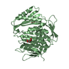
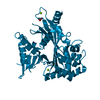
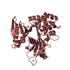
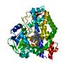
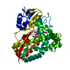
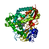
| Title | Structure of EspG Protein | ||||||
 Components Components | LEE-encoded effector EspG | ||||||
 Keywords Keywords | SIGNALING PROTEIN / VirA fold / virulence factor / PAK recruitment and activation / p21 activated kinase | ||||||
| Function / homology |  Function and homology information Function and homology informationcysteine-type endopeptidase activity / proteolysis / extracellular region Similarity search - Function | ||||||
| Biological species |  | ||||||
| Method |  X-RAY DIFFRACTION / SYNCHROTRON / SAD / Resolution: 1.596 Å X-RAY DIFFRACTION / SYNCHROTRON / SAD / Resolution: 1.596 Å | ||||||
 Authors Authors | Spiller, B.W. / Germane, K.L. | ||||||
 Citation Citation | Journal: Biochemistry / Year: 2011 Title: Structural and Functional Studies Indicate That the EPEC Effector, EspG, Directly Binds p21-Activated Kinase. Authors: Germane, K.L. / Spiller, B.W. | ||||||
| History |
|
- Structure visualization
| Structure viewer | Molecule: MolmilJmol/JSmol |
|---|
- Downloads & links
-Download
| PDBx/mmCIF format | 3q1c.cif.gz | 157.3 KB | Display | PDBx/mmCIF format |
|---|---|---|---|---|
| PDB format | pdb3q1c.ent.gz | 123.3 KB | Display | PDB format |
| PDBx/mmJSON format | 3q1c.json.gz | Tree view | PDBx/mmJSON format | |
| Others |  Other downloads Other downloads |
-Validation report
| Arichive directory | https://data.pdbj.org/pub/pdb/validation_reports/q1/3q1cftp://data.pdbj.org/pub/pdb/validation_reports/q1/3q1c | HTTPS FTP |
|---|
-Related structure data
| Similar structure data |
|---|


-Links
 PDBj
PDBj- Assembly
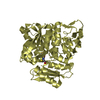
| Deposited unit | 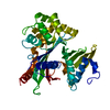
| ||||||||
|---|---|---|---|---|---|---|---|---|---|
| 1 |
| ||||||||
| Unit cell |
|
-Components
| #1: Protein | Mass: 39879.578 Da / Num. of mol.: 1 / Fragment: UNP residues 44-398 Source method: isolated from a genetically manipulated source Source: (gene. exp.) Plasmid details: An N-terminal 6His tag was cleaved with Thrombin. Plasmid: pET28a / Production host: |
|---|---|
| #2: Water | ChemComp-HOH /  Mass: 18.015 Da / Num. of mol.: 446 / Source method: isolated from a natural source / Formula: H2O Mass: 18.015 Da / Num. of mol.: 446 / Source method: isolated from a natural source / Formula: H2O |
-Experimental details
-Experiment
| Experiment | Method: X-RAY DIFFRACTION / Number of used crystals: 1 |
|---|
- Sample preparation
| Crystal | Density Matthews: 3.12 Å3/Da / Density % sol: 60.52 % |
|---|---|
| Crystal grow | Method: vapor diffusion, hanging drop Details: 2.0-11.0% PEG8000, 100 mM Bis-Tris, pH 6.25-7.5, VAPOR DIFFUSION, HANGING DROP, temperature range 268-291K PH range: 6.25-7.5 |
-Data collection
| Diffraction | Mean temperature: 100 K | |||||||||||
|---|---|---|---|---|---|---|---|---|---|---|---|---|
| Diffraction source | Source: SYNCHROTRON / Site: APS  / Beamline: 24-ID-C / Wavelength: 0.9795 / Beamline: 24-ID-C / Wavelength: 0.9795 | |||||||||||
| Detector | Type: ADSC QUANTUM 315 / Detector: CCD / Date: Jul 16, 2009 | |||||||||||
| Radiation | Protocol: SINGLE WAVELENGTH / Monochromatic (M) / Laue (L): M / Scattering type: x-ray | |||||||||||
| Radiation wavelength | Wavelength: 0.9795 Å / Relative weight: 1 | |||||||||||
| Reflection | Resolution: 1.595→32.958 Å / Num. obs: 60098 / Observed criterion σ(F): 0 / Observed criterion σ(I): 0 / Redundancy: 4.8 % / Biso Wilson estimate: 21.2 Å2 / Rmerge(I) obs: 0.035 / Net I/σ(I): 32 | |||||||||||
| Reflection shell |
|
- Processing
| Software |
| |||||||||||||||||||||||||||||||||||||||||||||||||||||||||||||||||||||||||||||||||||||||||||||||||||||||||
|---|---|---|---|---|---|---|---|---|---|---|---|---|---|---|---|---|---|---|---|---|---|---|---|---|---|---|---|---|---|---|---|---|---|---|---|---|---|---|---|---|---|---|---|---|---|---|---|---|---|---|---|---|---|---|---|---|---|---|---|---|---|---|---|---|---|---|---|---|---|---|---|---|---|---|---|---|---|---|---|---|---|---|---|---|---|---|---|---|---|---|---|---|---|---|---|---|---|---|---|---|---|---|---|---|---|---|
| Refinement | Method to determine structure: SAD / Resolution: 1.596→32.093 Å / SU ML: 0.18 / σ(F): 0 / Stereochemistry target values: ML
| |||||||||||||||||||||||||||||||||||||||||||||||||||||||||||||||||||||||||||||||||||||||||||||||||||||||||
| Solvent computation | Shrinkage radii: 0.61 Å / VDW probe radii: 0.9 Å / Solvent model: FLAT BULK SOLVENT MODEL / Bsol: 56.988 Å2 / ksol: 0.394 e/Å3 | |||||||||||||||||||||||||||||||||||||||||||||||||||||||||||||||||||||||||||||||||||||||||||||||||||||||||
| Displacement parameters |
| |||||||||||||||||||||||||||||||||||||||||||||||||||||||||||||||||||||||||||||||||||||||||||||||||||||||||
| Refine analyze | Luzzati sigma a obs: 0.18 Å | |||||||||||||||||||||||||||||||||||||||||||||||||||||||||||||||||||||||||||||||||||||||||||||||||||||||||
| Refinement step | Cycle: LAST / Resolution: 1.596→32.093 Å
| |||||||||||||||||||||||||||||||||||||||||||||||||||||||||||||||||||||||||||||||||||||||||||||||||||||||||
| Refine LS restraints |
| |||||||||||||||||||||||||||||||||||||||||||||||||||||||||||||||||||||||||||||||||||||||||||||||||||||||||
| LS refinement shell |
|
