 Movie
Movie Controller

[English] 日本語
 Yorodumi
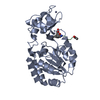
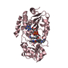
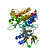
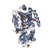
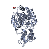
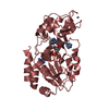
- PDB-5bwn: Crystal Structure of SIRT3 with a H3K9 Peptide Containing a Myris... -
+ Open data

- Basic information
| Entry | Database: PDB / ID: 5bwn | ||||||
|---|---|---|---|---|---|---|---|
| Title | Crystal Structure of SIRT3 with a H3K9 Peptide Containing a Myristoyl Lysine | ||||||
 Components Components |
| ||||||
 Keywords Keywords | PEPTIDE/HYDROLASE / Hydrolase / PEPTIDE-HYDROLASE complex | ||||||
| Function / homology |  Function and homology information Function and homology informationpositive regulation of catalase activity / positive regulation of superoxide dismutase activity / NAD-dependent protein lysine delactylase activity / positive regulation of ceramide biosynthetic process / peptidyl-lysine deacetylation / Maturation of TCA enzymes and regulation of TCA cycle / protein acetyllysine N-acetyltransferase / NAD-dependent protein lysine deacetylase activity / protein deacetylation / histone deacetylase activity, NAD-dependent ...positive regulation of catalase activity / positive regulation of superoxide dismutase activity / NAD-dependent protein lysine delactylase activity / positive regulation of ceramide biosynthetic process / peptidyl-lysine deacetylation / Maturation of TCA enzymes and regulation of TCA cycle / protein acetyllysine N-acetyltransferase / NAD-dependent protein lysine deacetylase activity / protein deacetylation / histone deacetylase activity, NAD-dependent / positive regulation of oxidative phosphorylation / Regulation of FOXO transcriptional activity by acetylation / protein lysine deacetylase activity / nucleosomal DNA binding / cellular response to stress / negative regulation of reactive oxygen species metabolic process / NAD+ binding / FOXO-mediated transcription of oxidative stress, metabolic and neuronal genes / Mitochondrial unfolded protein response (UPRmt) / Transferases; Acyltransferases; Transferring groups other than aminoacyl groups / aerobic respiration / Transcriptional activation of mitochondrial biogenesis / euchromatin / negative regulation of ERK1 and ERK2 cascade / positive regulation of insulin secretion / structural constituent of chromatin / nucleosome / positive regulation of cell growth / sequence-specific DNA binding / mitochondrial matrix / protein heterodimerization activity / enzyme binding / protein-containing complex / mitochondrion / zinc ion binding / nucleoplasm / nucleus Similarity search - Function | ||||||
| Biological species |  Homo sapiens (human) Homo sapiens (human)synthetic construct (others) | ||||||
| Method |  X-RAY DIFFRACTION / SYNCHROTRON / MOLECULAR REPLACEMENT / molecular replacement / Resolution: 1.942 Å X-RAY DIFFRACTION / SYNCHROTRON / MOLECULAR REPLACEMENT / molecular replacement / Resolution: 1.942 Å | ||||||
 Authors Authors | Gai, W. / Liu, D. | ||||||
 Citation Citation | Journal: Febs Lett. / Year: 2016 Title: Crystal structures of SIRT3 reveal that the alpha 2-alpha 3 loop and alpha 3-helix affect the interaction with long-chain acyl lysine. Authors: Gai, W. / Li, H. / Jiang, H. / Long, Y. / Liu, D. | ||||||
| History |
|
- Structure visualization
| Structure viewer | Molecule: MolmilJmol/JSmol |
|---|
- Downloads & links
-Download
| PDBx/mmCIF format | 5bwn.cif.gz | 121.9 KB | Display | PDBx/mmCIF format |
|---|---|---|---|---|
| PDB format | pdb5bwn.ent.gz | 93.7 KB | Display | PDB format |
| PDBx/mmJSON format | 5bwn.json.gz | Tree view | PDBx/mmJSON format | |
| Others |  Other downloads Other downloads |
-Validation report
| Arichive directory | https://data.pdbj.org/pub/pdb/validation_reports/bw/5bwnftp://data.pdbj.org/pub/pdb/validation_reports/bw/5bwn | HTTPS FTP |
|---|
-Related structure data
| Related structure data |  5bwoC 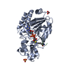 3gltS  5bwp 5bwq C: citing same article ( S: Starting model for refinement |
|---|---|
| Similar structure data |


-Links
 PDBj
PDBj


- Assembly
| Deposited unit | 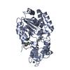
| ||||||||
|---|---|---|---|---|---|---|---|---|---|
| 1 |
| ||||||||
| Unit cell |
|
-Components
| #1: Protein/peptide | Mass: 1302.542 Da / Num. of mol.: 1 / Source method: obtained synthetically / Source: (synth.) synthetic construct (others) / References: UniProt: Q6NXT2*PLUS |
|---|---|
| #2: Protein | Mass: 34402.445 Da / Num. of mol.: 1 / Fragment: UNP RESIDUES 118-399 Source method: isolated from a genetically manipulated source Source: (gene. exp.) Homo sapiens (human) / Gene: SIRT3, SIR2L3 / Production host:  References: UniProt: Q9NTG7, Hydrolases; Acting on carbon-nitrogen bonds, other than peptide bonds; In linear amides |
| #3: Chemical | ChemComp-ZN /   Mass: 65.409 Da / Num. of mol.: 1 / Source method: obtained synthetically / Formula: Zn Mass: 65.409 Da / Num. of mol.: 1 / Source method: obtained synthetically / Formula: Zn |
| #4: Water | ChemComp-HOH /  Mass: 18.015 Da / Num. of mol.: 111 / Source method: isolated from a natural source / Formula: H2O Mass: 18.015 Da / Num. of mol.: 111 / Source method: isolated from a natural source / Formula: H2O |
-Experimental details
-Experiment
| Experiment | Method: X-RAY DIFFRACTION / Number of used crystals: 1 |
|---|
- Sample preparation
| Crystal | Density Matthews: 1.98 Å3/Da / Density % sol: 37.89 % |
|---|---|
| Crystal grow | Temperature: 293 K / Method: vapor diffusion, hanging drop / pH: 7 Details: 0.2M Ammonium tartrate dibasic (pH 7.0), 20% w/v PEG 3350, 0.01M Urea |
-Data collection
| Diffraction | Mean temperature: 100 K |
|---|---|
| Diffraction source | Source: SYNCHROTRON / Site: SSRF  / Beamline: BL17U / Wavelength: 0.9792 Å / Beamline: BL17U / Wavelength: 0.9792 Å |
| Detector | Type: MARMOSAIC 225 mm CCD / Detector: CCD / Date: Apr 21, 2015 / Details: M |
| Radiation | Protocol: SINGLE WAVELENGTH / Monochromatic (M) / Laue (L): M / Scattering type: x-ray |
| Radiation wavelength | Wavelength: 0.9792 Å / Relative weight: 1 |
| Reflection | Resolution: 1.94→50 Å / Num. obs: 21698 / % possible obs: 96 % / Redundancy: 11.98 % / Rmerge(I) obs: 0.077 / Net I/σ(I): 18.2 |
| Reflection shell | Resolution: 1.94→1.99 Å / Mean I/σ(I) obs: 5.2 / % possible all: 97.8 |
-Phasing
| Phasing | Method: molecular replacement |
|---|
- Processing
| Software |
| |||||||||||||||||||||||||||||||||||||||||||||||||||||||||||||||||||||||||||||||||||||||||||||||||||||||||
|---|---|---|---|---|---|---|---|---|---|---|---|---|---|---|---|---|---|---|---|---|---|---|---|---|---|---|---|---|---|---|---|---|---|---|---|---|---|---|---|---|---|---|---|---|---|---|---|---|---|---|---|---|---|---|---|---|---|---|---|---|---|---|---|---|---|---|---|---|---|---|---|---|---|---|---|---|---|---|---|---|---|---|---|---|---|---|---|---|---|---|---|---|---|---|---|---|---|---|---|---|---|---|---|---|---|---|
| Refinement | Method to determine structure: MOLECULAR REPLACEMENT Starting model: 3GLT Resolution: 1.942→41.279 Å / SU ML: 0.26 / Cross valid method: THROUGHOUT / σ(F): 1.36 / Phase error: 29.47 / Stereochemistry target values: ML
| |||||||||||||||||||||||||||||||||||||||||||||||||||||||||||||||||||||||||||||||||||||||||||||||||||||||||
| Solvent computation | Shrinkage radii: 0.9 Å / VDW probe radii: 1.11 Å / Solvent model: FLAT BULK SOLVENT MODEL | |||||||||||||||||||||||||||||||||||||||||||||||||||||||||||||||||||||||||||||||||||||||||||||||||||||||||
| Refinement step | Cycle: LAST / Resolution: 1.942→41.279 Å
| |||||||||||||||||||||||||||||||||||||||||||||||||||||||||||||||||||||||||||||||||||||||||||||||||||||||||
| Refine LS restraints |
| |||||||||||||||||||||||||||||||||||||||||||||||||||||||||||||||||||||||||||||||||||||||||||||||||||||||||
| LS refinement shell |
|
