 Movie
Movie Controller

[English] 日本語
 Yorodumi
- PDB-5bwl: Crystal Structure of SIRT5 in Complex with a Coumarin-Labelled Su... -
+ Open data

- Basic information
| Entry | Database: PDB / ID: 5bwl | ||||||
|---|---|---|---|---|---|---|---|
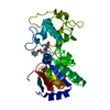
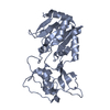
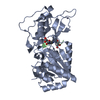
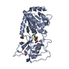
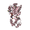
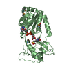
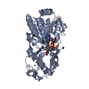
| Title | Crystal Structure of SIRT5 in Complex with a Coumarin-Labelled Succinyl Peptide | ||||||
 Components Components |
| ||||||
 Keywords Keywords | HYDROLASE | ||||||
| Function / homology |  Function and homology information Function and homology informationprotein demalonylation / protein deglutarylation / regulation of ketone biosynthetic process / peptidyl-lysine demalonylation / protein desuccinylation / peptidyl-lysine desuccinylation / protein-glutaryllysine deglutarylase activity / protein-malonyllysine demalonylase activity / protein-succinyllysine desuccinylase activity / protein deacetylation ...protein demalonylation / protein deglutarylation / regulation of ketone biosynthetic process / peptidyl-lysine demalonylation / protein desuccinylation / peptidyl-lysine desuccinylation / protein-glutaryllysine deglutarylase activity / protein-malonyllysine demalonylase activity / protein-succinyllysine desuccinylase activity / protein deacetylation / NAD-dependent protein lysine deacetylase activity / histone deacetylase activity, NAD-dependent / negative regulation of reactive oxygen species metabolic process / negative regulation of cardiac muscle cell apoptotic process / NAD+ binding / Transferases; Acyltransferases; Transferring groups other than aminoacyl groups / response to ischemia / mitochondrion organization / Transcriptional activation of mitochondrial biogenesis / response to nutrient levels / mitochondrial intermembrane space / mitochondrial inner membrane / mitochondrial matrix / mitochondrion / zinc ion binding / nucleus / cytosol Similarity search - Function | ||||||
| Biological species |  Homo sapiens (human) Homo sapiens (human)synthetic construct (others) | ||||||
| Method |  X-RAY DIFFRACTION / SYNCHROTRON / MOLECULAR REPLACEMENT / molecular replacement / Resolution: 1.548 Å X-RAY DIFFRACTION / SYNCHROTRON / MOLECULAR REPLACEMENT / molecular replacement / Resolution: 1.548 Å | ||||||
 Authors Authors | Gai, W. / Jiang, H. / Liu, D. | ||||||
 Citation Citation | Journal: To be published Title: Crystal Structure of SIRT5 in Complex with a Coumarin-Labelled Succinyl Peptide Authors: Gai, W. / Jiang, H. / Liu, D. | ||||||
| History |
|
- Structure visualization
| Structure viewer | Molecule: MolmilJmol/JSmol |
|---|
- Downloads & links
-Download
| PDBx/mmCIF format | 5bwl.cif.gz | 121.5 KB | Display | PDBx/mmCIF format |
|---|---|---|---|---|
| PDB format | pdb5bwl.ent.gz | 92.4 KB | Display | PDB format |
| PDBx/mmJSON format | 5bwl.json.gz | Tree view | PDBx/mmJSON format | |
| Others |  Other downloads Other downloads |
-Validation report
| Arichive directory | https://data.pdbj.org/pub/pdb/validation_reports/bw/5bwlftp://data.pdbj.org/pub/pdb/validation_reports/bw/5bwl | HTTPS FTP |
|---|
-Related structure data
| Related structure data | 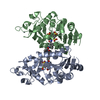 2nyrS  5bwp 5bwq S: Starting model for refinement |
|---|---|
| Similar structure data |


-Links
 PDBj
PDBj

- Assembly
| Deposited unit | 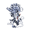
| ||||||||
|---|---|---|---|---|---|---|---|---|---|
| 1 |
| ||||||||
| Unit cell |
|
-Components
| #1: Protein | Mass: 31584.936 Da / Num. of mol.: 1 / Fragment: UNP RESIDUES 33-302 Source method: isolated from a genetically manipulated source Source: (gene. exp.) Homo sapiens (human) / Gene: SIRT5, SIR2L5 / Production host:  References: UniProt: Q9NXA8, Hydrolases; Acting on carbon-nitrogen bonds, other than peptide bonds; In linear amides |
|---|---|
| #2: Protein/peptide | Mass: 416.470 Da / Num. of mol.: 1 / Source method: obtained synthetically / Source: (synth.) synthetic construct (others) |
| #3: Chemical | ChemComp-ZN /   Mass: 65.409 Da / Num. of mol.: 1 / Source method: obtained synthetically / Formula: Zn Mass: 65.409 Da / Num. of mol.: 1 / Source method: obtained synthetically / Formula: Zn |
| #4: Chemical | ChemComp-MCM /   Mass: 175.184 Da / Num. of mol.: 1 / Source method: obtained synthetically / Formula: C10H9NO2 Mass: 175.184 Da / Num. of mol.: 1 / Source method: obtained synthetically / Formula: C10H9NO2 |
| #5: Water | ChemComp-HOH /  Mass: 18.015 Da / Num. of mol.: 376 / Source method: isolated from a natural source / Formula: H2O Mass: 18.015 Da / Num. of mol.: 376 / Source method: isolated from a natural source / Formula: H2O |
-Experimental details
-Experiment
| Experiment | Method: X-RAY DIFFRACTION / Number of used crystals: 1 |
|---|
- Sample preparation
| Crystal | Density Matthews: 2.49 Å3/Da / Density % sol: 50.52 % |
|---|---|
| Crystal grow | Temperature: 293 K / Method: vapor diffusion, hanging drop / pH: 6.5 / Details: 0.1M Bis-Tris (pH 6.5), 20% PEG 3350 |
-Data collection
| Diffraction | Mean temperature: 100 K |
|---|---|
| Diffraction source | Source: SYNCHROTRON / Site: SSRF  / Beamline: BL17U / Wavelength: 0.9792 Å / Beamline: BL17U / Wavelength: 0.9792 Å |
| Detector | Type: MARMOSAIC 225 mm CCD / Detector: CCD / Date: Apr 21, 2015 / Details: M |
| Radiation | Protocol: SINGLE WAVELENGTH / Monochromatic (M) / Laue (L): M / Scattering type: x-ray |
| Radiation wavelength | Wavelength: 0.9792 Å / Relative weight: 1 |
| Reflection | Resolution: 1.548→50 Å / Num. obs: 43833 / % possible obs: 97.7 % / Redundancy: 4.59 % / Rmerge(I) obs: 0.06 / Net I/σ(I): 25.3 |
| Reflection shell | Resolution: 1.55→1.58 Å / Redundancy: 3.7 % / Mean I/σ(I) obs: 6.9 / % possible all: 100 |
-Phasing
| Phasing | Method: molecular replacement |
|---|
- Processing
| Software |
| |||||||||||||||||||||||||||||||||||||||||||||||||||||||||||||||||||||||||||||||||||||||||||||||||||||||||
|---|---|---|---|---|---|---|---|---|---|---|---|---|---|---|---|---|---|---|---|---|---|---|---|---|---|---|---|---|---|---|---|---|---|---|---|---|---|---|---|---|---|---|---|---|---|---|---|---|---|---|---|---|---|---|---|---|---|---|---|---|---|---|---|---|---|---|---|---|---|---|---|---|---|---|---|---|---|---|---|---|---|---|---|---|---|---|---|---|---|---|---|---|---|---|---|---|---|---|---|---|---|---|---|---|---|---|
| Refinement | Method to determine structure: MOLECULAR REPLACEMENT Starting model: 2NYR Resolution: 1.548→35.071 Å / SU ML: 0.19 / Cross valid method: THROUGHOUT / σ(F): 1.34 / Phase error: 23.42 / Stereochemistry target values: ML
| |||||||||||||||||||||||||||||||||||||||||||||||||||||||||||||||||||||||||||||||||||||||||||||||||||||||||
| Solvent computation | Shrinkage radii: 0.9 Å / VDW probe radii: 1.11 Å / Solvent model: FLAT BULK SOLVENT MODEL | |||||||||||||||||||||||||||||||||||||||||||||||||||||||||||||||||||||||||||||||||||||||||||||||||||||||||
| Refinement step | Cycle: LAST / Resolution: 1.548→35.071 Å
| |||||||||||||||||||||||||||||||||||||||||||||||||||||||||||||||||||||||||||||||||||||||||||||||||||||||||
| Refine LS restraints |
| |||||||||||||||||||||||||||||||||||||||||||||||||||||||||||||||||||||||||||||||||||||||||||||||||||||||||
| LS refinement shell |
|
