 Movie
Movie Controller


+ Open data

- Basic information
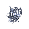
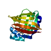
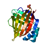
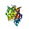
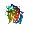
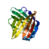
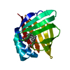
| Entry | Database: PDB / ID: 5bvt | ||||||
|---|---|---|---|---|---|---|---|
| Title | Palmitate-bound pFABP5 | ||||||
 Components Components | Epidermal fatty acid-binding protein | ||||||
 Keywords Keywords | LIPID BINDING PROTEIN / Fatty acid-binding protein / beta-barrel protein / Gentoo penguin (Pygoscelis papua) | ||||||
| Function / homology |  Function and homology information Function and homology informationlipid transport / postsynaptic density / lipid binding / extracellular region / nucleus / cytoplasm Similarity search - Function | ||||||
| Biological species |  Pygoscelis papua (Gentoo penguin) Pygoscelis papua (Gentoo penguin) | ||||||
| Method |  X-RAY DIFFRACTION / SYNCHROTRON / Resolution: 2.31 Å X-RAY DIFFRACTION / SYNCHROTRON / Resolution: 2.31 Å | ||||||
 Authors Authors | Lee, J.H. / Lee, C.W. / Do, H. | ||||||
 Citation Citation | Journal: Biochem.Biophys.Res.Commun. / Year: 2015 Title: Structural basis for the ligand-binding specificity of fatty acid-binding proteins (pFABP4 and pFABP5) in gentoo penguin Authors: Lee, C.W. / Kim, J.E. / Do, H. / Kim, R.O. / Lee, S.G. / Park, H.H. / Chang, J.H. / Yim, J.H. / Park, H. / Kim, I.C. / Lee, J.H. | ||||||
| History |
|
- Structure visualization
| Structure viewer | Molecule: MolmilJmol/JSmol |
|---|
- Downloads & links
-Download
| PDBx/mmCIF format | 5bvt.cif.gz | 41.7 KB | Display | PDBx/mmCIF format |
|---|---|---|---|---|
| PDB format | pdb5bvt.ent.gz | 27.5 KB | Display | PDB format |
| PDBx/mmJSON format | 5bvt.json.gz | Tree view | PDBx/mmJSON format | |
| Others |  Other downloads Other downloads |
-Validation report
| Arichive directory | https://data.pdbj.org/pub/pdb/validation_reports/bv/5bvtftp://data.pdbj.org/pub/pdb/validation_reports/bv/5bvt | HTTPS FTP |
|---|
-Related structure data


-Links
 PDBj
PDBj

- Assembly
| Deposited unit | 
| ||||||||
|---|---|---|---|---|---|---|---|---|---|
| 1 |
| ||||||||
| Unit cell |
|
-Components
| #1: Protein | Mass: 15127.494 Da / Num. of mol.: 1 Source method: isolated from a genetically manipulated source Source: (gene. exp.) Pygoscelis papua (Gentoo penguin) / Production host:  |
|---|---|
| #2: Chemical | ChemComp-PAM / 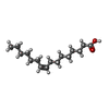  Mass: 254.408 Da / Num. of mol.: 1 / Source method: obtained synthetically / Formula: C16H30O2 Mass: 254.408 Da / Num. of mol.: 1 / Source method: obtained synthetically / Formula: C16H30O2 |
| #3: Water | ChemComp-HOH /  Mass: 18.015 Da / Num. of mol.: 56 / Source method: isolated from a natural source / Formula: H2O Mass: 18.015 Da / Num. of mol.: 56 / Source method: isolated from a natural source / Formula: H2O |
| Sequence details | A SEQUENCE DATABASE REFERENCE FOR THIS PROTEIN DOES NOT CURRENTLY EXIST. |
-Experimental details
-Experiment
| Experiment | Method: X-RAY DIFFRACTION / Number of used crystals: 1 |
|---|
- Sample preparation
| Crystal | Density Matthews: 1.93 Å3/Da / Density % sol: 36.33 % |
|---|---|
| Crystal grow | Temperature: 293 K / Method: vapor diffusion, hanging drop Details: 0.2 M magnesium chloride, 0.1 M Tris (pH 8.5), 30% (w/v) PEG 4000 |
-Data collection
| Diffraction | Mean temperature: 100 K |
|---|---|
| Diffraction source | Source: SYNCHROTRON / Site: PAL/PLS  / Beamline: 7A (6B, 6C1) / Wavelength: 0.9793 Å / Beamline: 7A (6B, 6C1) / Wavelength: 0.9793 Å |
| Detector | Type: ADSC QUANTUM 315r / Detector: CCD / Date: Dec 6, 2014 |
| Radiation | Protocol: SINGLE WAVELENGTH / Monochromatic (M) / Laue (L): M / Scattering type: x-ray |
| Radiation wavelength | Wavelength: 0.9793 Å / Relative weight: 1 |
| Reflection | Resolution: 2.3→50 Å / Num. obs: 4997 / % possible obs: 97.6 % / Redundancy: 7.5 % / Net I/σ(I): 61.3 |
- Processing
| Software |
| ||||||||||||||||||||||||||||||||||||||||||||||||||||||||||||||||||||||||||||||||||||||||||||||||||||||||||||||||||||||||||||||||||||||||||||||||||||||||||||||||||||||||||||||||||||||
|---|---|---|---|---|---|---|---|---|---|---|---|---|---|---|---|---|---|---|---|---|---|---|---|---|---|---|---|---|---|---|---|---|---|---|---|---|---|---|---|---|---|---|---|---|---|---|---|---|---|---|---|---|---|---|---|---|---|---|---|---|---|---|---|---|---|---|---|---|---|---|---|---|---|---|---|---|---|---|---|---|---|---|---|---|---|---|---|---|---|---|---|---|---|---|---|---|---|---|---|---|---|---|---|---|---|---|---|---|---|---|---|---|---|---|---|---|---|---|---|---|---|---|---|---|---|---|---|---|---|---|---|---|---|---|---|---|---|---|---|---|---|---|---|---|---|---|---|---|---|---|---|---|---|---|---|---|---|---|---|---|---|---|---|---|---|---|---|---|---|---|---|---|---|---|---|---|---|---|---|---|---|---|---|
| Refinement | Resolution: 2.31→32.89 Å / Cor.coef. Fo:Fc: 0.954 / Cor.coef. Fo:Fc free: 0.873 / SU B: 9.157 / SU ML: 0.222 / Cross valid method: THROUGHOUT / ESU R: 0.865 / ESU R Free: 0.31 / Stereochemistry target values: MAXIMUM LIKELIHOOD / Details: HYDROGENS HAVE BEEN ADDED IN THE RIDING POSITIONS
| ||||||||||||||||||||||||||||||||||||||||||||||||||||||||||||||||||||||||||||||||||||||||||||||||||||||||||||||||||||||||||||||||||||||||||||||||||||||||||||||||||||||||||||||||||||||
| Solvent computation | Ion probe radii: 0.8 Å / Shrinkage radii: 0.8 Å / VDW probe radii: 1.2 Å / Solvent model: MASK | ||||||||||||||||||||||||||||||||||||||||||||||||||||||||||||||||||||||||||||||||||||||||||||||||||||||||||||||||||||||||||||||||||||||||||||||||||||||||||||||||||||||||||||||||||||||
| Displacement parameters | Biso mean: 32.062 Å2
| ||||||||||||||||||||||||||||||||||||||||||||||||||||||||||||||||||||||||||||||||||||||||||||||||||||||||||||||||||||||||||||||||||||||||||||||||||||||||||||||||||||||||||||||||||||||
| Refinement step | Cycle: 1 / Resolution: 2.31→32.89 Å
| ||||||||||||||||||||||||||||||||||||||||||||||||||||||||||||||||||||||||||||||||||||||||||||||||||||||||||||||||||||||||||||||||||||||||||||||||||||||||||||||||||||||||||||||||||||||
| Refine LS restraints |
|