 Movie
Movie Controller

[English] 日本語
 Yorodumi
- PDB-1g74: Toward changing specificity: adipocyte lipid binding protein muta... -
+ Open data

- Basic information
| Entry | Database: PDB / ID: 1g74 | ||||||
|---|---|---|---|---|---|---|---|
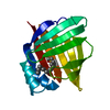
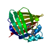
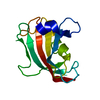
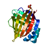
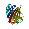
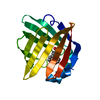
| Title | Toward changing specificity: adipocyte lipid binding protein mutant, oleic acid bound form | ||||||
 Components Components | ADIPOCYTE LIPID-BINDING PROTEIN | ||||||
 Keywords Keywords | LIPID BINDING PROTEIN / beta-barrel / fatty acid binding protein / protein engineering / fatty acid binding | ||||||
| Function / homology |  Function and homology information Function and homology informationlipase activator activity / Triglyceride catabolism / lipid droplet formation / triglyceride catabolic process / hormone receptor binding / long-chain fatty acid transmembrane transporter activity / long-chain fatty acid binding / cellular response to lithium ion / white fat cell differentiation / long-chain fatty acid transport ...lipase activator activity / Triglyceride catabolism / lipid droplet formation / triglyceride catabolic process / hormone receptor binding / long-chain fatty acid transmembrane transporter activity / long-chain fatty acid binding / cellular response to lithium ion / white fat cell differentiation / long-chain fatty acid transport / fatty acid transport / lipid droplet / brown fat cell differentiation / fatty acid binding / cholesterol homeostasis / response to bacterium / fatty acid metabolic process / cellular response to tumor necrosis factor / positive regulation of inflammatory response / positive regulation of cold-induced thermogenesis / negative regulation of DNA-templated transcription / positive regulation of cell population proliferation / nucleoplasm / nucleus / cytoplasm / cytosol Similarity search - Function | ||||||
| Biological species |  | ||||||
| Method |  X-RAY DIFFRACTION / isomorphous with apo EF-ALBP (PDB 1G7N) / Resolution: 1.7 Å X-RAY DIFFRACTION / isomorphous with apo EF-ALBP (PDB 1G7N) / Resolution: 1.7 Å | ||||||
 Authors Authors | Reese, A.J. / Banaszak, L.J. | ||||||
 Citation Citation | Journal: J.Lipid Res. / Year: 2004 Title: Specificity determinants for lipids bound to beta-barrel proteins. Authors: Reese, A.J. / Banaszak, L.J. | ||||||
| History |
|
- Structure visualization
| Structure viewer | Molecule: MolmilJmol/JSmol |
|---|
- Downloads & links
-Download
| PDBx/mmCIF format | 1g74.cif.gz | 43.6 KB | Display | PDBx/mmCIF format |
|---|---|---|---|---|
| PDB format | pdb1g74.ent.gz | 30.1 KB | Display | PDB format |
| PDBx/mmJSON format | 1g74.json.gz | Tree view | PDBx/mmJSON format | |
| Others |  Other downloads Other downloads |
-Validation report
| Arichive directory | https://data.pdbj.org/pub/pdb/validation_reports/g7/1g74ftp://data.pdbj.org/pub/pdb/validation_reports/g7/1g74 | HTTPS FTP |
|---|
-Related structure data
| Related structure data |  1g7nSC S: Starting model for refinement C: citing same article ( |
|---|---|
| Similar structure data |


-Links
 PDBj
PDBj


- Assembly
| Deposited unit | 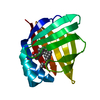
| ||||||||
|---|---|---|---|---|---|---|---|---|---|
| 1 |
| ||||||||
| Unit cell |
| ||||||||
| Components on special symmetry positions |
|

-Components
| #1: Protein | Mass: 14525.663 Da / Num. of mol.: 1 / Mutation: I73E,A75V,D77G Source method: isolated from a genetically manipulated source Source: (gene. exp.)  |
|---|---|
| #2: Chemical | ChemComp-PO4 /   Mass: 94.971 Da / Num. of mol.: 1 / Source method: obtained synthetically / Formula: PO4 Mass: 94.971 Da / Num. of mol.: 1 / Source method: obtained synthetically / Formula: PO4 |
| #3: Chemical | ChemComp-OLA /   Mass: 282.461 Da / Num. of mol.: 1 / Source method: obtained synthetically / Formula: C18H34O2 Mass: 282.461 Da / Num. of mol.: 1 / Source method: obtained synthetically / Formula: C18H34O2 |
| #4: Water | ChemComp-HOH /  Mass: 18.015 Da / Num. of mol.: 119 / Source method: isolated from a natural source / Formula: H2O Mass: 18.015 Da / Num. of mol.: 119 / Source method: isolated from a natural source / Formula: H2O |
-Experimental details
-Experiment
| Experiment | Method: X-RAY DIFFRACTION / Number of used crystals: 1 |
|---|
- Sample preparation
| Crystal | Density Matthews: 2.1 Å3/Da / Density % sol: 41.49 % | |||||||||||||||||||||||||||||||||||||||||||||||||
|---|---|---|---|---|---|---|---|---|---|---|---|---|---|---|---|---|---|---|---|---|---|---|---|---|---|---|---|---|---|---|---|---|---|---|---|---|---|---|---|---|---|---|---|---|---|---|---|---|---|---|
| Crystal grow | Temperature: 291 K / Method: vapor diffusion, hanging drop / pH: 6.8 Details: sodium/potassium phosphate, pH 6.8, VAPOR DIFFUSION, HANGING DROP, temperature 291K | |||||||||||||||||||||||||||||||||||||||||||||||||
| Crystal grow | *PLUS pH: 7.2 / Method: vapor diffusion, hanging drop | |||||||||||||||||||||||||||||||||||||||||||||||||
| Components of the solutions | *PLUS
|
-Data collection
| Diffraction | Mean temperature: 113 K |
|---|---|
| Diffraction source | Source: ROTATING ANODE / Type: RIGAKU / Wavelength: 1.5418 Å |
| Detector | Type: RIGAKU RAXIS IV / Detector: IMAGE PLATE / Date: Jul 13, 2000 / Details: mirrors |
| Radiation | Monochromator: graphite / Protocol: SINGLE WAVELENGTH / Monochromatic (M) / Laue (L): M / Scattering type: x-ray |
| Radiation wavelength | Wavelength: 1.5418 Å / Relative weight: 1 |
| Reflection | Resolution: 1.71→27 Å / Num. obs: 12433 / % possible obs: 93.9 % / Observed criterion σ(F): 3 / Observed criterion σ(I): 3 / Redundancy: 3.31 % / Rmerge(I) obs: 0.046 / Net I/σ(I): 16.4 |
| Reflection shell | Resolution: 1.71→1.77 Å / Redundancy: 3.37 % / Rmerge(I) obs: 0.304 / % possible all: 90.1 |
| Reflection | *PLUS Highest resolution: 1.7 Å / Lowest resolution: 25 Å / Num. obs: 13235 / Redundancy: 3.19 % / Num. measured all: 42285 |
- Processing
| Software |
| ||||||||||||||||||||
|---|---|---|---|---|---|---|---|---|---|---|---|---|---|---|---|---|---|---|---|---|---|
| Refinement | Method to determine structure: isomorphous with apo EF-ALBP (PDB 1G7N) Starting model: EF-ALBP (PDB 1G7N) Resolution: 1.7→25 Å / σ(F): 0 / σ(I): 0 / Stereochemistry target values: Engh & Huber Details: The following amino acids have alternative conformations: Cys1, Lys9, Lys21, Glu22, Val44, Thr74, Glu116, Ser124, Glu129, and Oleate132
| ||||||||||||||||||||
| Refinement step | Cycle: LAST / Resolution: 1.7→25 Å
| ||||||||||||||||||||
| LS refinement shell | Resolution: 1.71→1.77 Å /
| ||||||||||||||||||||
| Refinement | *PLUS Rfactor Rfree: 0.237 / Rfactor Rwork: 0.208 | ||||||||||||||||||||
| Solvent computation | *PLUS | ||||||||||||||||||||
| Displacement parameters | *PLUS | ||||||||||||||||||||
| Refine LS restraints | *PLUS
|
