 Movie
Movie Controller


+ Open data

- Basic information
| Entry | Database: PDB / ID: 5bsa | ||||||
|---|---|---|---|---|---|---|---|
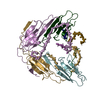
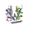
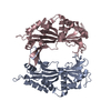
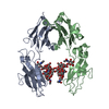
| Title | Structure of histone H3/H4 in complex with Spt2 | ||||||
 Components Components |
| ||||||
 Keywords Keywords | TRANSCRIPTION REGULATOR / chaperone / transcription | ||||||
| Function / homology |  Function and homology information Function and homology informationhistone chaperone activity / RNA polymerase I core binding / transcription by RNA polymerase I / structural constituent of chromatin / nucleosome / nucleosome assembly / heterochromatin formation / histone binding / protein heterodimerization activity / regulation of DNA-templated transcription ...histone chaperone activity / RNA polymerase I core binding / transcription by RNA polymerase I / structural constituent of chromatin / nucleosome / nucleosome assembly / heterochromatin formation / histone binding / protein heterodimerization activity / regulation of DNA-templated transcription / nucleolus / DNA binding / nucleoplasm / nucleus Similarity search - Function | ||||||
| Biological species |  Homo sapiens (human) Homo sapiens (human) | ||||||
| Method |  X-RAY DIFFRACTION / SYNCHROTRON / MOLECULAR REPLACEMENT / Resolution: 4.611 Å X-RAY DIFFRACTION / SYNCHROTRON / MOLECULAR REPLACEMENT / Resolution: 4.611 Å | ||||||
 Authors Authors | Chen, S. / Patel, D.J. | ||||||
 Citation Citation | Journal: Genes Dev. / Year: 2015 Title: Structure-function studies of histone H3/H4 tetramer maintenance during transcription by chaperone Spt2. Authors: Chen, S. / Rufiange, A. / Huang, H. / Rajashankar, K.R. / Nourani, A. / Patel, D.J. | ||||||
| History |
|
- Structure visualization
| Structure viewer | Molecule: MolmilJmol/JSmol |
|---|
- Downloads & links
-Download
| PDBx/mmCIF format | 5bsa.cif.gz | 82.4 KB | Display | PDBx/mmCIF format |
|---|---|---|---|---|
| PDB format | pdb5bsa.ent.gz | 58.2 KB | Display | PDB format |
| PDBx/mmJSON format | 5bsa.json.gz | Tree view | PDBx/mmJSON format | |
| Others |  Other downloads Other downloads |
-Validation report
| Arichive directory | https://data.pdbj.org/pub/pdb/validation_reports/bs/5bsaftp://data.pdbj.org/pub/pdb/validation_reports/bs/5bsa | HTTPS FTP |
|---|
-Related structure data
| Related structure data |  5bs7C 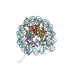 1aoiS C: citing same article ( S: Starting model for refinement |
|---|---|
| Similar structure data |


-Links
 PDBj
PDBj


- Assembly
| Deposited unit | 
| ||||||||
|---|---|---|---|---|---|---|---|---|---|
| 1 |
| ||||||||
| Unit cell |
|
-Components
| #1: Protein | Mass: 12673.827 Da / Num. of mol.: 2 / Fragment: residues 27-136 Source method: isolated from a genetically manipulated source Source: (gene. exp.)  #2: Protein | Mass: 11263.231 Da / Num. of mol.: 2 Source method: isolated from a genetically manipulated source Source: (gene. exp.) #3: Protein | Mass: 14651.086 Da / Num. of mol.: 2 / Fragment: residues 571-685 Source method: isolated from a genetically manipulated source Source: (gene. exp.) Homo sapiens (human) / Gene: SPTY2D1 / Production host: Has protein modification | Y | |
|---|
-Experimental details
-Experiment
| Experiment | Method: X-RAY DIFFRACTION |
|---|
- Sample preparation
| Crystal | Density Matthews: 2.97 Å3/Da / Density % sol: 58.57 % |
|---|---|
| Crystal grow | Temperature: 293 K / Method: vapor diffusion / pH: 7.5 Details: 0.02 M NaCl, 0.2 M HEPES 7.5, 1.6 M ammonium sulfate |
-Data collection
| Diffraction | Mean temperature: 100 K |
|---|---|
| Diffraction source | Source: SYNCHROTRON / Site: APS  / Beamline: 24-ID-E / Wavelength: 0.9792 Å / Beamline: 24-ID-E / Wavelength: 0.9792 Å |
| Detector | Type: ADSC QUANTUM 315 / Detector: CCD / Date: Jun 11, 2013 |
| Radiation | Protocol: SINGLE WAVELENGTH / Monochromatic (M) / Laue (L): M / Scattering type: x-ray |
| Radiation wavelength | Wavelength: 0.9792 Å / Relative weight: 1 |
| Reflection | Resolution: 4.6→50 Å / Num. obs: 5704 / % possible obs: 98.7 % / Redundancy: 11.4 % / Rmerge(I) obs: 0.066 / Net I/σ(I): 22.3 |
| Reflection shell | Resolution: 4.6→4.68 Å / Redundancy: 7.8 % / Mean I/σ(I) obs: 1.3 / % possible all: 88.6 |
- Processing
| Software |
| ||||||||||||||||||||||||||||
|---|---|---|---|---|---|---|---|---|---|---|---|---|---|---|---|---|---|---|---|---|---|---|---|---|---|---|---|---|---|
| Refinement | Method to determine structure: MOLECULAR REPLACEMENT Starting model: 1AOI Resolution: 4.611→49.115 Å / SU ML: 0.89 / Cross valid method: FREE R-VALUE / σ(F): 1.35 / Phase error: 38.13 / Stereochemistry target values: ML
| ||||||||||||||||||||||||||||
| Solvent computation | Shrinkage radii: 0.9 Å / VDW probe radii: 1.11 Å / Solvent model: FLAT BULK SOLVENT MODEL | ||||||||||||||||||||||||||||
| Refinement step | Cycle: LAST / Resolution: 4.611→49.115 Å
| ||||||||||||||||||||||||||||
| Refine LS restraints |
| ||||||||||||||||||||||||||||
| LS refinement shell |
|
