Resolution: 2.2→48.2 Å / Cor.coef. Fo:Fc: 0.933 / Cor.coef. Fo:Fc free: 0.905 / SU B: 6.717 / SU ML: 0.172 / Cross valid method: THROUGHOUT / ESU R: 0.272 / ESU R Free: 0.226 / Stereochemistry target values: MAXIMUM LIKELIHOOD / Details: HYDROGENS HAVE BEEN ADDED IN THE RIDING POSITIONS
Rfactor
Num. reflection
% reflection
Selection details
Rfree
0.27495
1250
5.1 %
RANDOM
Rwork
0.22393
-
-
-
obs
0.22656
23338
99.8 %
-
Solvent computation
Ion probe radii: 0.8 Å / Shrinkage radii: 0.8 Å / VDW probe radii: 1.2 Å / Solvent model: MASK
 Movie
Movie Controller


 Open data
 Basic information
 Components
 Keywords
 Function and homology information
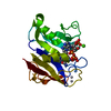
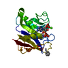
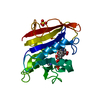
 Clostridium perfringens (bacteria)
 X-RAY DIFFRACTION /
  Authors
 Japan, 1items
  Citation
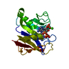
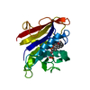
 Structure visualization
 Downloads & links
 Other downloads


 PDBj
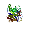
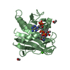
PDBj Assembly
 Mass: 18.015 Da / Num. of mol.: 107 / Source method: isolated from a natural source / Formula: H2O
 Sample preparation
 Processing