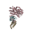
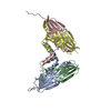
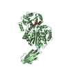
Component-ID: _ / Ens-ID: 1 / Beg auth comp-ID: GLY / Beg label comp-ID: GLY / End auth comp-ID: SER / End label comp-ID: SER / Refine code: _ / Auth seq-ID: 38 - 301 / Label seq-ID: 52 - 315
Dom-ID
Auth asym-ID
Label asym-ID
1
A
A
2
B
B
-
Components
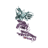
#1: Protein
MatrixproteinVP40 / Membrane-associated protein VP40
Mass: 36017.359 Da / Num. of mol.: 2 Source method: isolated from a genetically manipulated source Source: (gene. exp.) Lake Victoria marburgvirus (strain Musoke-80) Strain: Musoke-80 / Gene: VP40 / Production host: Escherichia coli (E. coli) / References: UniProt: P35260
Resolution: 2.81→50 Å / Num. obs: 25037 / % possible obs: 99.5 % / Redundancy: 4 % / Net I/σ(I): 6.9
-
Processing
Software
Name
Version
Classification
REFMAC
5.8.0073
refinement
HKL-2000
datareduction
HKL-2000
datascaling
Auto-Rickshaw
phasing
Refinement
Method to determine structure: MAD / Resolution: 2.81→29.67 Å / Cor.coef. Fo:Fc: 0.931 / Cor.coef. Fo:Fc free: 0.879 / SU B: 35.489 / SU ML: 0.317 / Cross valid method: THROUGHOUT / ESU R Free: 0.373 / Stereochemistry target values: MAXIMUM LIKELIHOOD / Details: HYDROGENS HAVE BEEN ADDED IN THE RIDING POSITIONS
Rfactor
Num. reflection
% reflection
Selection details
Rfree
0.25686
828
5.1 %
RANDOM
Rwork
0.20182
-
-
-
obs
0.20459
15475
99 %
-
Solvent computation
Ion probe radii: 0.8 Å / Shrinkage radii: 0.8 Å / VDW probe radii: 1.2 Å / Solvent model: MASK
 Movie
Movie Controller


 Open data
 Basic information
 Components
 Keywords
 Function and homology information
 Lake Victoria marburgvirus
 X-RAY DIFFRACTION /
  Authors
 Citation
 Structure visualization
 Downloads & links
 Other downloads
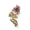

 PDBj
PDBj


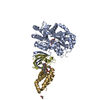
 Assembly


 Mass: 46.068 Da / Num. of mol.: 1 / Source method: obtained synthetically / Formula: C2H6O
 Mass: 18.015 Da / Num. of mol.: 35 / Source method: isolated from a natural source / Formula: H2O
 Sample preparation
 / Beamline: 23-ID-B / Wavelength: 0.979, 0.980
 Processing