 Movie
Movie Controller

[English] 日本語
 Yorodumi
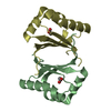
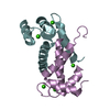
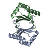
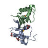
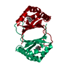
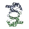
- PDB-5b09: Polyketide cyclase OAC from Cannabis sativa bound with Olivetolic acid -
+ Open data

- Basic information
| Entry | Database: PDB / ID: 5b09 | ||||||
|---|---|---|---|---|---|---|---|
| Title | Polyketide cyclase OAC from Cannabis sativa bound with Olivetolic acid | ||||||
 Components Components | Olivetolic acid cyclase | ||||||
 Keywords Keywords | LYASE / Cannabis sativa / plant polyketide cyclase | ||||||
| Function / homology |  Function and homology information Function and homology informationolivetolic acid cyclase / olivetolic acid biosynthetic process / cannabinoid biosynthetic process / pollen tube adhesion / cyclase activity / terpenoid biosynthetic process / lyase activity / metal ion binding / cytoplasm Similarity search - Function | ||||||
| Biological species |  Cannabis sativa (plant) Cannabis sativa (plant) | ||||||
| Method |  X-RAY DIFFRACTION / SYNCHROTRON / MOLECULAR REPLACEMENT / molecular replacement / Resolution: 1.7 Å X-RAY DIFFRACTION / SYNCHROTRON / MOLECULAR REPLACEMENT / molecular replacement / Resolution: 1.7 Å | ||||||
 Authors Authors | Yang, X. / Matsui, T. / Mori, T. / Abe, I. / Morita, H. | ||||||
 Citation Citation | Journal: Febs J. / Year: 2016 Title: Structural basis for olivetolic acid formation by a polyketide cyclase from Cannabis sativa Authors: Yang, X. / Matsui, T. / Kodama, T. / Mori, T. / Zhou, X. / Taura, F. / Noguchi, H. / Abe, I. / Morita, H. | ||||||
| History |
|
- Structure visualization
| Structure viewer | Molecule: MolmilJmol/JSmol |
|---|
- Downloads & links
-Download
| PDBx/mmCIF format | 5b09.cif.gz | 36.3 KB | Display | PDBx/mmCIF format |
|---|---|---|---|---|
| PDB format | pdb5b09.ent.gz | 22.6 KB | Display | PDB format |
| PDBx/mmJSON format | 5b09.json.gz | Tree view | PDBx/mmJSON format | |
| Others |  Other downloads Other downloads |
-Validation report
| Arichive directory | https://data.pdbj.org/pub/pdb/validation_reports/b0/5b09ftp://data.pdbj.org/pub/pdb/validation_reports/b0/5b09 | HTTPS FTP |
|---|
-Related structure data
| Related structure data |  5b08SC  5b0aC  5b0bC  5b0cC  5b0dC  5b0eC  5b0fC  5b0gC S: Starting model for refinement C: citing same article ( |
|---|---|
| Similar structure data |


-Links
 PDBj
PDBj- Assembly
| Deposited unit | 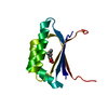
| ||||||||
|---|---|---|---|---|---|---|---|---|---|
| 1 | 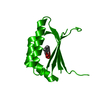
| ||||||||
| Unit cell |
|
-Components
| #1: Protein | Mass: 12230.011 Da / Num. of mol.: 1 Source method: isolated from a genetically manipulated source Source: (gene. exp.) Cannabis sativa (plant) / Gene: OAC / Plasmid: pQE-80L / Production host:  |
|---|---|
| #2: Chemical | ChemComp-4MX / 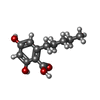  Mass: 224.253 Da / Num. of mol.: 1 / Source method: obtained synthetically / Formula: C12H16O4 Mass: 224.253 Da / Num. of mol.: 1 / Source method: obtained synthetically / Formula: C12H16O4 |
| #3: Water | ChemComp-HOH /  Mass: 18.015 Da / Num. of mol.: 56 / Source method: isolated from a natural source / Formula: H2O Mass: 18.015 Da / Num. of mol.: 56 / Source method: isolated from a natural source / Formula: H2O |
-Experimental details
-Experiment
| Experiment | Method: X-RAY DIFFRACTION / Number of used crystals: 1 |
|---|
- Sample preparation
| Crystal | Density Matthews: 1.89 Å3/Da / Density % sol: 34.98 % |
|---|---|
| Crystal grow | Temperature: 278 K / Method: vapor diffusion, sitting drop / pH: 8.8 Details: 100mM Tris - HCl pH 8.8, 25%(w/v) PEG6000, 50mM olivetolic acid, 5%(v/v) methanol |
-Data collection
| Diffraction | Mean temperature: 100 K |
|---|---|
| Diffraction source | Source: SYNCHROTRON / Site: Photon Factory  / Beamline: AR-NE3A / Wavelength: 1 Å / Beamline: AR-NE3A / Wavelength: 1 Å |
| Detector | Type: ADSC QUANTUM 270 / Detector: CCD / Date: Jun 14, 2014 |
| Radiation | Monochromator: Si(1 1 1) / Protocol: SINGLE WAVELENGTH / Monochromatic (M) / Laue (L): M / Scattering type: x-ray |
| Radiation wavelength | Wavelength: 1 Å / Relative weight: 1 |
| Reflection | Resolution: 1.7→50 Å / Num. obs: 10098 / % possible obs: 98.5 % / Redundancy: 6.8 % / Rmerge(I) obs: 0.041 / Net I/σ(I): 27.9 |
| Reflection shell | Resolution: 1.7→1.8 Å / Redundancy: 4.7 % / Rmerge(I) obs: 0.31 / Mean I/σ(I) obs: 5.1 / % possible all: 92.3 |
-Phasing
| Phasing | Method: molecular replacement | |||||||||
|---|---|---|---|---|---|---|---|---|---|---|
| Phasing MR |
|
- Processing
| Software |
| |||||||||||||||||||||||||||||||||||
|---|---|---|---|---|---|---|---|---|---|---|---|---|---|---|---|---|---|---|---|---|---|---|---|---|---|---|---|---|---|---|---|---|---|---|---|---|
| Refinement | Method to determine structure: MOLECULAR REPLACEMENT Starting model: 5B08 Resolution: 1.7→32.112 Å / SU ML: 0.18 / Cross valid method: FREE R-VALUE / σ(F): 1.39 / Phase error: 25.52 / Stereochemistry target values: ML
| |||||||||||||||||||||||||||||||||||
| Solvent computation | Shrinkage radii: 0.9 Å / VDW probe radii: 1.11 Å / Solvent model: FLAT BULK SOLVENT MODEL | |||||||||||||||||||||||||||||||||||
| Displacement parameters | Biso max: 60.58 Å2 / Biso mean: 24.0696 Å2 / Biso min: 11.01 Å2 | |||||||||||||||||||||||||||||||||||
| Refinement step | Cycle: final / Resolution: 1.7→32.112 Å
| |||||||||||||||||||||||||||||||||||
| Refine LS restraints |
| |||||||||||||||||||||||||||||||||||
| LS refinement shell | Refine-ID: X-RAY DIFFRACTION / Total num. of bins used: 4
|
