 Movie
Movie Controller


+ Open data

- Basic information
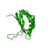
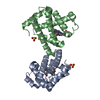
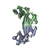
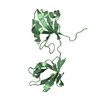
| Entry | Database: PDB / ID: 1tuv | ||||||
|---|---|---|---|---|---|---|---|
| Title | Crystal structure of YgiN in complex with menadione | ||||||
 Components Components | Protein ygiN | ||||||
 Keywords Keywords | UNKNOWN FUNCTION / menadione oxidase / monooxygenase / co-crystal with natural product / ferredoxin fold | ||||||
| Function / homology |  Function and homology information Function and homology informationOxidoreductases / response to acidic pH / catalytic activity / oxidoreductase activity / protein homodimerization activity / cytosol Similarity search - Function | ||||||
| Biological species |  | ||||||
| Method |  X-RAY DIFFRACTION / SYNCHROTRON / direct input of native model / Resolution: 1.7 Å X-RAY DIFFRACTION / SYNCHROTRON / direct input of native model / Resolution: 1.7 Å | ||||||
 Authors Authors | Adams, M.A. / Jia, Z. | ||||||
 Citation Citation | Journal: J.Biol.Chem. / Year: 2005 Title: Structural and Biochemical Evidence for an Enzymatic Quinone Redox Cycle in Escherichia coli: IDENTIFICATION OF A NOVEL QUINOL MONOOXYGENASE Authors: Adams, M.A. / Jia, Z. | ||||||
| History |
|
- Structure visualization
| Structure viewer | Molecule: MolmilJmol/JSmol |
|---|
- Downloads & links
-Download
| PDBx/mmCIF format | 1tuv.cif.gz | 38.4 KB | Display | PDBx/mmCIF format |
|---|---|---|---|---|
| PDB format | pdb1tuv.ent.gz | 26.5 KB | Display | PDB format |
| PDBx/mmJSON format | 1tuv.json.gz | Tree view | PDBx/mmJSON format | |
| Others |  Other downloads Other downloads |
-Validation report
| Arichive directory | https://data.pdbj.org/pub/pdb/validation_reports/tu/1tuvftp://data.pdbj.org/pub/pdb/validation_reports/tu/1tuv | HTTPS FTP |
|---|
-Related structure data


-Links
 PDBj
PDBj- Assembly
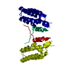
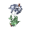
| Deposited unit | 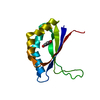
| ||||||||
|---|---|---|---|---|---|---|---|---|---|
| 1 | 
| ||||||||
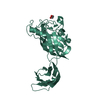
| 2 | 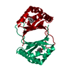
| ||||||||
| 3 |
| ||||||||
| Unit cell |
| ||||||||
| Components on special symmetry positions |
|
-Components
| #1: Protein | Mass: 12775.716 Da / Num. of mol.: 1 Source method: isolated from a genetically manipulated source Source: (gene. exp.) |
|---|---|
| #2: Chemical | ChemComp-VK3 /   Mass: 172.180 Da / Num. of mol.: 1 / Source method: obtained synthetically / Formula: C11H8O2 Mass: 172.180 Da / Num. of mol.: 1 / Source method: obtained synthetically / Formula: C11H8O2 |
| #3: Water | ChemComp-HOH /  Mass: 18.015 Da / Num. of mol.: 181 / Source method: isolated from a natural source / Formula: H2O Mass: 18.015 Da / Num. of mol.: 181 / Source method: isolated from a natural source / Formula: H2O |
-Experimental details
-Experiment
| Experiment | Method: X-RAY DIFFRACTION / Number of used crystals: 1 |
|---|
- Sample preparation
| Crystal | Density Matthews: 3.24 Å3/Da / Density % sol: 61.7 % |
|---|---|
| Crystal grow | Temperature: 298 K / Method: vapor diffusion, hanging drop / pH: 5.6 Details: sodium citrate, ammonium sulfate, sodium/potassium tartrate, pH 5.6, VAPOR DIFFUSION, HANGING DROP, temperature 298K |
-Data collection
| Diffraction | Mean temperature: 100 K |
|---|---|
| Diffraction source | Source: SYNCHROTRON / Site: NSLS  / Beamline: X6A / Wavelength: 0.9788 Å / Beamline: X6A / Wavelength: 0.9788 Å |
| Detector | Type: ADSC QUANTUM 210 / Detector: CCD / Date: Apr 25, 2004 |
| Radiation | Monochromator: Si(111) channel cut monochromator / Protocol: SINGLE WAVELENGTH / Monochromatic (M) / Laue (L): M / Scattering type: x-ray |
| Radiation wavelength | Wavelength: 0.9788 Å / Relative weight: 1 |
| Reflection | Resolution: 1.69→50 Å / Num. all: 19783 / Num. obs: 19783 / % possible obs: 100 % / Observed criterion σ(F): 2 / Observed criterion σ(I): 2 / Redundancy: 8 % / Rmerge(I) obs: 0.07 / Rsym value: 0.07 / Net I/σ(I): 50 |
| Reflection shell | Resolution: 1.69→1.77 Å / % possible all: 99 |
- Processing
| Software |
| ||||||||||||||||||||||||||||||||||||||||||||||||||||||||||||||||||||||||||||||||||||||||||||||||||||
|---|---|---|---|---|---|---|---|---|---|---|---|---|---|---|---|---|---|---|---|---|---|---|---|---|---|---|---|---|---|---|---|---|---|---|---|---|---|---|---|---|---|---|---|---|---|---|---|---|---|---|---|---|---|---|---|---|---|---|---|---|---|---|---|---|---|---|---|---|---|---|---|---|---|---|---|---|---|---|---|---|---|---|---|---|---|---|---|---|---|---|---|---|---|---|---|---|---|---|---|---|---|
| Refinement | Method to determine structure: direct input of native model / Resolution: 1.7→25.65 Å / Cor.coef. Fo:Fc: 0.958 / Cor.coef. Fo:Fc free: 0.928 / SU B: 1.913 / SU ML: 0.062 / Cross valid method: THROUGHOUT / σ(F): 0 / ESU R: 0.09 / ESU R Free: 0.097 / Stereochemistry target values: MAXIMUM LIKELIHOOD / Details: HYDROGENS HAVE BEEN ADDED IN THE RIDING POSITIONS
| ||||||||||||||||||||||||||||||||||||||||||||||||||||||||||||||||||||||||||||||||||||||||||||||||||||
| Solvent computation | Ion probe radii: 0.8 Å / Shrinkage radii: 0.8 Å / VDW probe radii: 1.4 Å / Solvent model: BABINET MODEL WITH MASK | ||||||||||||||||||||||||||||||||||||||||||||||||||||||||||||||||||||||||||||||||||||||||||||||||||||
| Displacement parameters | Biso mean: 22.765 Å2 | ||||||||||||||||||||||||||||||||||||||||||||||||||||||||||||||||||||||||||||||||||||||||||||||||||||
| Refinement step | Cycle: LAST / Resolution: 1.7→25.65 Å
| ||||||||||||||||||||||||||||||||||||||||||||||||||||||||||||||||||||||||||||||||||||||||||||||||||||
| Refine LS restraints |
| ||||||||||||||||||||||||||||||||||||||||||||||||||||||||||||||||||||||||||||||||||||||||||||||||||||
| LS refinement shell | Resolution: 1.701→1.745 Å / Total num. of bins used: 20 /
|
