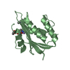
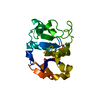
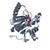
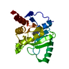
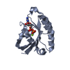
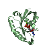
[English] 日本語
Yorodumi
- PDB-4aii: Crystal structure of the rat REM2 GTPase - G domain bound to GDP -

+
Open data


ID or keywords:

Loading...

-
Basic information

Entry
Database: PDB / ID: 4aii
TitleCrystal structure of the rat REM2 GTPase - G domain bound to GDP
ComponentsGTP-BINDING PROTEIN REM 2
KeywordsGTP-BINDING PROTEIN / RGK PROTEINS
Function / homology
Function and homology information


calcium channel regulator activity / GTPase activity / GTP binding / signal transduction / membrane / plasma membrane
Similarity search - Function
Sigma-54 interaction domain, ATP-binding site 1 / Small GTPase, Ras-type / small GTPase Ras family profile. / Small GTPase / Ras family / P-loop containing nucleotide triphosphate hydrolases / P-loop containing nucleoside triphosphate hydrolase / Rossmann fold / 3-Layer(aba) Sandwich / Alpha Beta
Similarity search - Domain/homology
GUANOSINE-5'-DIPHOSPHATE / GTP-binding protein REM 2
Similarity search - Component
Biological speciesRATTUS NORVEGICUS (Norway rat)
MethodX-RAY DIFFRACTION / SYNCHROTRON / MOLECULAR REPLACEMENT / Resolution: 2.66 Å
AuthorsReymond, P. / Coquard, A. / Chenon, M. / Zeghouf, M. / El Marjou, A. / Thompson, A. / Menetrey, J.
CitationJournal: Acta Crystallogr.,Sect.F / Year: 2012
Title: Structure of the Gdp-Bound G Domain of the Rgk Protein Rem2.
Authors: Reymond, P. / Coquard, A. / Chenon, M. / Zeghouf, M. / El Marjou, A. / Thompson, A. / Menetrey, J.
History
DepositionFeb 10, 2012Deposition site: PDBE / Processing site: PDBE
Revision 1.0Jun 13, 2012Provider: repository / Type: Initial release
Revision 1.1Jun 20, 2012Group: Other
Revision 1.2Dec 20, 2023Group: Data collection / Database references ...Data collection / Database references / Derived calculations / Other / Refinement description
Category: chem_comp_atom / chem_comp_bond ...chem_comp_atom / chem_comp_bond / database_2 / pdbx_database_status / pdbx_initial_refinement_model / pdbx_struct_conn_angle / struct_conn / struct_site
Item: _database_2.pdbx_DOI / _database_2.pdbx_database_accession ..._database_2.pdbx_DOI / _database_2.pdbx_database_accession / _pdbx_database_status.status_code_sf / _pdbx_struct_conn_angle.ptnr1_auth_comp_id / _pdbx_struct_conn_angle.ptnr1_auth_seq_id / _pdbx_struct_conn_angle.ptnr1_label_asym_id / _pdbx_struct_conn_angle.ptnr1_label_atom_id / _pdbx_struct_conn_angle.ptnr1_label_comp_id / _pdbx_struct_conn_angle.ptnr1_label_seq_id / _pdbx_struct_conn_angle.ptnr3_auth_comp_id / _pdbx_struct_conn_angle.ptnr3_auth_seq_id / _pdbx_struct_conn_angle.ptnr3_label_asym_id / _pdbx_struct_conn_angle.ptnr3_label_atom_id / _pdbx_struct_conn_angle.ptnr3_label_comp_id / _pdbx_struct_conn_angle.ptnr3_label_seq_id / _pdbx_struct_conn_angle.value / _struct_conn.pdbx_dist_value / _struct_conn.ptnr1_auth_comp_id / _struct_conn.ptnr1_auth_seq_id / _struct_conn.ptnr1_label_asym_id / _struct_conn.ptnr1_label_atom_id / _struct_conn.ptnr1_label_comp_id / _struct_conn.ptnr1_label_seq_id / _struct_conn.ptnr2_auth_comp_id / _struct_conn.ptnr2_auth_seq_id / _struct_conn.ptnr2_label_asym_id / _struct_conn.ptnr2_label_atom_id / _struct_conn.ptnr2_label_comp_id / _struct_conn.ptnr2_label_seq_id / _struct_site.pdbx_auth_asym_id / _struct_site.pdbx_auth_comp_id / _struct_site.pdbx_auth_seq_id

-
Structure visualization

Structure viewerMolecule:
MolmilJmol/JSmol

Downloads & links

-
Assembly

Deposited unit
A: GTP-BINDING PROTEIN REM 2
B: GTP-BINDING PROTEIN REM 2
hetero molecules


Theoretical massNumber of molelcules
Total (without water)41,2426
Polymers40,3072
Non-polymers9354
Water21612
1
A: GTP-BINDING PROTEIN REM 2
hetero molecules


Theoretical massNumber of molelcules
Total (without water)20,6213
Polymers20,1541
Non-polymers4682
Water181
TypeNameSymmetry operationNumber
identity operation1_555x,y,z1
MethodPISA
2
B: GTP-BINDING PROTEIN REM 2
hetero molecules


Theoretical massNumber of molelcules
Total (without water)20,6213
Polymers20,1541
Non-polymers4682
Water181
TypeNameSymmetry operationNumber
identity operation1_555x,y,z1
MethodPISA
Unit cell
Length a, b, c (Å)50.916, 59.374, 57.297
Angle α, β, γ (deg.)90.00, 101.51, 90.00
Int Tables number4
Space group name H-MP1211
Noncrystallographic symmetry (NCS)NCS domain:
IDEns-IDDetails
11A
21B

NCS domain segments:
Dom-IDComponent-IDEns-IDRefine codeAuth asym-IDAuth seq-ID
1113A113 - 136
2113B113 - 136
1213A150 - 172
2213B150 - 172
1313A190 - 282
2313B190 - 282
1411A301 - 306
2411B301 - 306

-
Components

#1: Protein GTP-BINDING PROTEIN REM 2 / RAD AND GEM-LIKE GTP-BINDING PROTEIN 2


Mass: 20153.689 Da / Num. of mol.: 2 / Fragment: G DOMAIN, RESIDUES 113-283
Source method: isolated from a genetically manipulated source
Source: (gene. exp.) RATTUS NORVEGICUS (Norway rat) / Production host: ESCHERICHIA COLI (E. coli) / Strain (production host): BL21 / References: UniProt: Q9WTY2
#2: Chemical ChemComp-MG / MAGNESIUM ION


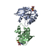
Mass: 24.305 Da / Num. of mol.: 2 / Source method: obtained synthetically / Formula: Mg
#3: Chemical ChemComp-GDP / GUANOSINE-5'-DIPHOSPHATE


Type: RNA linking / Mass: 443.201 Da / Num. of mol.: 2 / Source method: obtained synthetically / Formula: C10H15N5O11P2 / Comment: GDP, energy-carrying molecule*YM
#4: Water ChemComp-HOH / water


Mass: 18.015 Da / Num. of mol.: 12 / Source method: isolated from a natural source / Formula: H2O

-
Experimental details

-
Experiment

ExperimentMethod: X-RAY DIFFRACTION

-
Sample preparation

CrystalDensity Matthews: 2.11 Å3/Da / Density % sol: 41.58 % / Description: NONE
Crystal growDetails: 13.5 % PEG 4000, 10% ISOPROPANOL, 100 MM HEPES SODIUM SALT PH 7.4

-
Data collection

DiffractionMean temperature: 287 K
Diffraction sourceSource: SYNCHROTRON / Site: SOLEIL / Beamline: PROXIMA 1 / Wavelength: 0.98011
DetectorType: DECTRIS PILATUS 6M / Detector: PIXEL / Date: Oct 6, 2011
RadiationProtocol: SINGLE WAVELENGTH / Monochromatic (M) / Laue (L): M / Scattering type: x-ray
Radiation wavelengthWavelength: 0.98011 Å / Relative weight: 1
ReflectionResolution: 2.66→41.65 Å / Num. obs: 9524 / % possible obs: 98.8 % / Observed criterion σ(I): 2 / Redundancy: 4 % / Rmerge(I) obs: 0.11 / Net I/σ(I): 5.7
Reflection shellResolution: 2.66→2.8 Å / Rmerge(I) obs: 0.41 / Mean I/σ(I) obs: 2 / % possible all: 98.8

-
Processing

Software
NameVersionClassification
REFMAC5.5.0109refinement
XDSdata reduction
XDSdata scaling
PHASERphasing
RefinementMethod to determine structure: MOLECULAR REPLACEMENT
Starting model: PDB ENTRY 3CBQ
Resolution: 2.66→41.65 Å / Cor.coef. Fo:Fc: 0.916 / Cor.coef. Fo:Fc free: 0.832 / SU B: 14.91 / SU ML: 0.32 / Cross valid method: THROUGHOUT / ESU R: 3.941 / ESU R Free: 0.439 / Stereochemistry target values: MAXIMUM LIKELIHOOD
Details: HYDROGENS HAVE BEEN ADDED IN THE RIDING POSITIONS. RESIDUES 138-149 AND 171-182 ARE DISORDERED
RfactorNum. reflection% reflectionSelection details
Rfree0.32436 448 4.7 %RANDOM
Rwork0.24329 ---
obs0.24693 9160 98.36 %-
Solvent computationIon probe radii: 0.8 Å / Shrinkage radii: 0.8 Å / VDW probe radii: 1.4 Å / Solvent model: MASK
Displacement parametersBiso mean: 35.842 Å2
Baniso -1Baniso -2Baniso -3
1--0.77 Å20 Å20.64 Å2
2---5.06 Å20 Å2
3---6.09 Å2
Refinement stepCycle: LAST / Resolution: 2.66→41.65 Å
ProteinNucleic acidLigandSolventTotal
Num. atoms2174 0 58 12 2244
Refine LS restraints
Refine-IDTypeDev idealDev ideal targetNumber
X-RAY DIFFRACTIONr_bond_refined_d0.0140.0212298
X-RAY DIFFRACTIONr_bond_other_d
X-RAY DIFFRACTIONr_angle_refined_deg1.6331.9863115
X-RAY DIFFRACTIONr_angle_other_deg
X-RAY DIFFRACTIONr_dihedral_angle_1_deg6.5835288
X-RAY DIFFRACTIONr_dihedral_angle_2_deg34.75821.15895
X-RAY DIFFRACTIONr_dihedral_angle_3_deg17.51515372
X-RAY DIFFRACTIONr_dihedral_angle_4_deg19.8421526
X-RAY DIFFRACTIONr_chiral_restr0.0930.2366
X-RAY DIFFRACTIONr_gen_planes_refined0.0060.021682
X-RAY DIFFRACTIONr_gen_planes_other
X-RAY DIFFRACTIONr_nbd_refined
X-RAY DIFFRACTIONr_nbd_other
X-RAY DIFFRACTIONr_nbtor_refined
X-RAY DIFFRACTIONr_nbtor_other
X-RAY DIFFRACTIONr_xyhbond_nbd_refined
X-RAY DIFFRACTIONr_xyhbond_nbd_other
X-RAY DIFFRACTIONr_metal_ion_refined
X-RAY DIFFRACTIONr_metal_ion_other
X-RAY DIFFRACTIONr_symmetry_vdw_refined
X-RAY DIFFRACTIONr_symmetry_vdw_other
X-RAY DIFFRACTIONr_symmetry_hbond_refined
X-RAY DIFFRACTIONr_symmetry_hbond_other
X-RAY DIFFRACTIONr_symmetry_metal_ion_refined
X-RAY DIFFRACTIONr_symmetry_metal_ion_other
X-RAY DIFFRACTIONr_mcbond_it0.5751.51431
X-RAY DIFFRACTIONr_mcbond_other
X-RAY DIFFRACTIONr_mcangle_it1.11622277
X-RAY DIFFRACTIONr_mcangle_other
X-RAY DIFFRACTIONr_scbond_it1.9573867
X-RAY DIFFRACTIONr_scbond_other
X-RAY DIFFRACTIONr_scangle_it3.2754.5836
X-RAY DIFFRACTIONr_scangle_other
X-RAY DIFFRACTIONr_long_range_B_refined
X-RAY DIFFRACTIONr_long_range_B_other
X-RAY DIFFRACTIONr_rigid_bond_restr
X-RAY DIFFRACTIONr_sphericity_free
X-RAY DIFFRACTIONr_sphericity_bonded
Refine LS restraints NCS

Ens-ID: 1 / Refine-ID: X-RAY DIFFRACTION

Dom-IDAuth asym-IDNumberTypeRms dev position (Å)Weight position
1A582tight positional0.030.05
2B582tight positional0.030.05
1A489loose positional0.045
2B489loose positional0.045
1A582tight thermal0.320.5
2B582tight thermal0.320.5
1A489loose thermal0.6510
2B489loose thermal0.6510
LS refinement shellResolution: 2.66→2.729 Å / Total num. of bins used: 20
RfactorNum. reflection% reflection
Rfree0.334 42 -
Rwork0.329 667 -
obs--98.34 %

+
About Yorodumi

-
News

-
Feb 9, 2022. New format data for meta-information of EMDB entries

New format data for meta-information of EMDB entries

  • Version 3 of the EMDB header file is now the official format.
  • The previous official version 1.9 will be removed from the archive.

Related info.:EMDB header

External links:wwPDB to switch to version 3 of the EMDB data model

-
Aug 12, 2020. Covid-19 info

Covid-19 info

URL: https://pdbjlvh1.pdbj.org/emnavi/covid19.php

New page: Covid-19 featured information page in EM Navigator.

Related info.:Covid-19 info / Mar 5, 2020. Novel coronavirus structure data

+
Mar 5, 2020. Novel coronavirus structure data

Novel coronavirus structure data

Related info.:Yorodumi Speices / Aug 12, 2020. Covid-19 info

External links:COVID-19 featured content - PDBj / Molecule of the Month (242):Coronavirus Proteases

+
Jan 31, 2019. EMDB accession codes are about to change! (news from PDBe EMDB page)

EMDB accession codes are about to change! (news from PDBe EMDB page)

  • The allocation of 4 digits for EMDB accession codes will soon come to an end. Whilst these codes will remain in use, new EMDB accession codes will include an additional digit and will expand incrementally as the available range of codes is exhausted. The current 4-digit format prefixed with “EMD-” (i.e. EMD-XXXX) will advance to a 5-digit format (i.e. EMD-XXXXX), and so on. It is currently estimated that the 4-digit codes will be depleted around Spring 2019, at which point the 5-digit format will come into force.
  • The EM Navigator/Yorodumi systems omit the EMD- prefix.

Related info.:Q: What is EMD? / ID/Accession-code notation in Yorodumi/EM Navigator

External links:EMDB Accession Codes are Changing Soon! / Contact to PDBj

+
Jul 12, 2017. Major update of PDB

Major update of PDB

  • wwPDB released updated PDB data conforming to the new PDBx/mmCIF dictionary.
  • This is a major update changing the version number from 4 to 5, and with Remediation, in which all the entries are updated.
  • In this update, many items about electron microscopy experimental information are reorganized (e.g. em_software).
  • Now, EM Navigator and Yorodumi are based on the updated data.

External links:wwPDB Remediation / Enriched Model Files Conforming to OneDep Data Standards Now Available in the PDB FTP Archive

-
Yorodumi

Thousand views of thousand structures

  • Yorodumi is a browser for structure data from EMDB, PDB, SASBDB, etc.
  • This page is also the successor to EM Navigator detail page, and also detail information page/front-end page for Omokage search.
  • The word "yorodu" (or yorozu) is an old Japanese word meaning "ten thousand". "mi" (miru) is to see.

Related info.:EMDB / PDB / SASBDB / Comparison of 3 databanks / Yorodumi Search / Aug 31, 2016. New EM Navigator & Yorodumi / Yorodumi Papers / Jmol/JSmol / Function and homology information / Changes in new EM Navigator and Yorodumi

Read more