 Movie
Movie Controller


+ Open data

- Basic information
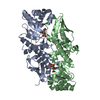
| Entry | Database: PDB / ID: 5ayx | |||||||||
|---|---|---|---|---|---|---|---|---|---|---|
| Title | Crystal structure of Human Quinolinate Phosphoribosyltransferase | |||||||||
 Components Components | Nicotinate-nucleotide pyrophosphorylase [carboxylating] | |||||||||
 Keywords Keywords | TRANSFERASE / QUINOLINATE PHOSPHORIBOSYLTRANSFERASE / NAD BIOSYNTHESIS / NADC | |||||||||
| Function / homology |  Function and homology information Function and homology informationquinolinate catabolic process / nicotinate-nucleotide diphosphorylase (carboxylating) / nicotinate-nucleotide diphosphorylase (carboxylating) activity / 'de novo' NAD+ biosynthetic process from L-tryptophan / NAD+ biosynthetic process / Nicotinate metabolism / catalytic complex / extracellular exosome / identical protein binding / cytosol / cytoplasm Similarity search - Function | |||||||||
| Biological species |  Homo sapiens (human) Homo sapiens (human) | |||||||||
| Method |  X-RAY DIFFRACTION / SYNCHROTRON / MOLECULAR REPLACEMENT / Resolution: 2.8 Å X-RAY DIFFRACTION / SYNCHROTRON / MOLECULAR REPLACEMENT / Resolution: 2.8 Å | |||||||||
 Authors Authors | Kang, G.B. / Kim, M.-K. / Im, Y.J. / Lee, J.H. / Youn, H.-S. / An, J.Y. / Lee, J.-G. / Fukuoka, S.-I. / Eom, S.H. | |||||||||
 Citation Citation | Journal: Sci Rep / Year: 2016 Title: Structural Insights into the Quaternary Catalytic Mechanism of Hexameric Human Quinolinate Phosphoribosyltransferase, a Key Enzyme in de novo NAD Biosynthesis Authors: Youn, H.-S. / Kim, T.G. / Kim, M.-K. / Kang, G.B. / Kang, J.Y. / Lee, J.-G. / An, J.Y. / Park, K.R. / Lee, Y. / Im, Y.J. / Lee, J.H. / Eom, S.H. | |||||||||
| History |
|
- Structure visualization
| Structure viewer | Molecule: MolmilJmol/JSmol |
|---|
- Downloads & links
-Download
| PDBx/mmCIF format | 5ayx.cif.gz | 310.6 KB | Display | PDBx/mmCIF format |
|---|---|---|---|---|
| PDB format | pdb5ayx.ent.gz | 254 KB | Display | PDB format |
| PDBx/mmJSON format | 5ayx.json.gz | Tree view | PDBx/mmJSON format | |
| Others |  Other downloads Other downloads |
-Validation report
| Arichive directory | https://data.pdbj.org/pub/pdb/validation_reports/ay/5ayxftp://data.pdbj.org/pub/pdb/validation_reports/ay/5ayx | HTTPS FTP |
|---|
-Related structure data
| Related structure data |  5ayyC  5ayzC  2jbmS S: Starting model for refinement C: citing same article ( |
|---|---|
| Similar structure data |


-Links
 PDBj
PDBj- Assembly
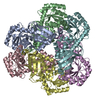
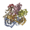
| Deposited unit | 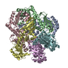
| ||||||||
|---|---|---|---|---|---|---|---|---|---|
| 1 |
| ||||||||
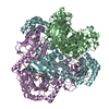
| Unit cell |
|
-Components
| #1: Protein | Mass: 31946.672 Da / Num. of mol.: 6 Source method: isolated from a genetically manipulated source Source: (gene. exp.) Homo sapiens (human) / Gene: QPRT / Plasmid: PET21A / Production host:  References: UniProt: Q15274, nicotinate-nucleotide diphosphorylase (carboxylating) #2: Water | ChemComp-HOH / |  Mass: 18.015 Da / Num. of mol.: 19 / Source method: isolated from a natural source / Formula: H2O Mass: 18.015 Da / Num. of mol.: 19 / Source method: isolated from a natural source / Formula: H2O |
|---|
-Experimental details
-Experiment
| Experiment | Method: X-RAY DIFFRACTION / Number of used crystals: 1 |
|---|
- Sample preparation
| Crystal | Density Matthews: 2.45 Å3/Da / Density % sol: 49.88 % |
|---|---|
| Crystal grow | Temperature: 294 K / Method: vapor diffusion / pH: 5 Details: 100MM MES-NAOH (PH 5.0), 7-15% (W/V) PEG-MME 2000, 10MM KSCN, VAPOR DIFFUSION, HANGING DROP, TEMPERATURE 294K |
-Data collection
| Diffraction | Mean temperature: 100 K |
|---|---|
| Diffraction source | Source: SYNCHROTRON / Site: Photon Factory  / Beamline: AR-NW12A / Wavelength: 1 Å / Beamline: AR-NW12A / Wavelength: 1 Å |
| Detector | Type: ADSC QUANTUM 315 / Detector: CCD / Date: Jun 30, 2004 |
| Radiation | Monochromator: DOUBLE CRYSTAL MONOCHROMATOR / Protocol: SINGLE WAVELENGTH / Monochromatic (M) / Laue (L): M / Scattering type: x-ray |
| Radiation wavelength | Wavelength: 1 Å / Relative weight: 1 |
| Reflection | Resolution: 2.8→50 Å / Num. obs: 42775 / % possible obs: 96.7 % / Observed criterion σ(I): 0 / Redundancy: 4.6 % / Net I/σ(I): 18.3 |
| Reflection shell | Resolution: 2.8→2.9 Å / % possible all: 94.2 |
- Processing
| Software |
| ||||||||||||||||||
|---|---|---|---|---|---|---|---|---|---|---|---|---|---|---|---|---|---|---|---|
| Refinement | Method to determine structure: MOLECULAR REPLACEMENT Starting model: PDB ENTRY 2JBM Resolution: 2.8→48 Å / Cross valid method: FREE R-VALUE
| ||||||||||||||||||
| Displacement parameters | Biso mean: 64 Å2
| ||||||||||||||||||
| Refinement step | Cycle: LAST / Resolution: 2.8→48 Å
| ||||||||||||||||||
| LS refinement shell | Resolution: 2.8→2.9 Å / Rfactor Rfree: 0.356 / Rfactor Rwork: 0.259 |
