 Movie
Movie Controller

[English] 日本語
 Yorodumi
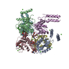
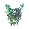
- PDB-5axn: Crystal structure of Thg1 like protein (TLP) with tRNA(Phe) and GDPNP -
+ Open data

- Basic information
| Entry | Database: PDB / ID: 5axn | ||||||
|---|---|---|---|---|---|---|---|
| Title | Crystal structure of Thg1 like protein (TLP) with tRNA(Phe) and GDPNP | ||||||
 Components Components |
| ||||||
 Keywords Keywords | Transferase/RNA / Transferase / Transferase-RNA complex | ||||||
| Function / homology |  Function and homology information Function and homology informationtRNA guanylyltransferase activity / tRNA modification / GTP binding / magnesium ion binding Similarity search - Function | ||||||
| Biological species |  Methanosarcina acetivorans (archaea) Methanosarcina acetivorans (archaea) | ||||||
| Method |  X-RAY DIFFRACTION / SYNCHROTRON / MOLECULAR REPLACEMENT / Resolution: 2.703 Å X-RAY DIFFRACTION / SYNCHROTRON / MOLECULAR REPLACEMENT / Resolution: 2.703 Å | ||||||
 Authors Authors | Kimura, S. / Suzuki, T. / Yu, J. / Kato, K. / Yao, M. | ||||||
 Citation Citation | Journal: Sci Adv / Year: 2016 Title: Template-dependent nucleotide addition in the reverse (3'-5') direction by Thg1-like protein Authors: Kimura, S. / Suzuki, T. / Chen, M. / Kato, K. / Yu, J. / Nakamura, A. / Tanaka, I. / Yao, M. | ||||||
| History |
|
- Structure visualization
| Structure viewer | Molecule: MolmilJmol/JSmol |
|---|
- Downloads & links
-Download
| PDBx/mmCIF format | 5axn.cif.gz | 150.7 KB | Display | PDBx/mmCIF format |
|---|---|---|---|---|
| PDB format | pdb5axn.ent.gz | 111.7 KB | Display | PDB format |
| PDBx/mmJSON format | 5axn.json.gz | Tree view | PDBx/mmJSON format | |
| Others |  Other downloads Other downloads |
-Validation report
| Summary document | 5axn_validation.pdf.gz | 1.1 MB | Display | wwPDB validaton report |
|---|---|---|---|---|
| Full document | 5axn_full_validation.pdf.gz | 1.1 MB | Display | |
| Data in XML | 5axn_validation.xml.gz | 21.2 KB | Display | |
| Data in CIF | 5axn_validation.cif.gz | 28.5 KB | Display | |
| Arichive directory | https://data.pdbj.org/pub/pdb/validation_reports/ax/5axnftp://data.pdbj.org/pub/pdb/validation_reports/ax/5axn | HTTPS FTP |
-Related structure data
| Related structure data | 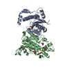 5axkC  5axlC  5axmC 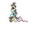 1ehzS 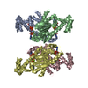 3wbzS C: citing same article ( S: Starting model for refinement |
|---|---|
| Similar structure data |


-Links
 PDBj
PDBj


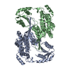
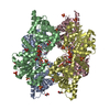
- Assembly
| Deposited unit | 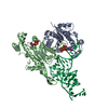
| ||||||||
|---|---|---|---|---|---|---|---|---|---|
| 1 | 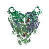
| ||||||||
| Unit cell |
|
-Components
| #1: Protein | Mass: 29325.727 Da / Num. of mol.: 2 Source method: isolated from a genetically manipulated source Source: (gene. exp.) Methanosarcina acetivorans (archaea) / Plasmid: pET26b / Production host:  #2: RNA chain | | Mass: 24332.340 Da / Num. of mol.: 1 / Source method: obtained synthetically / Source: (synth.) #3: Chemical | ChemComp-MG /   Mass: 24.305 Da / Num. of mol.: 5 / Source method: obtained synthetically / Formula: Mg Mass: 24.305 Da / Num. of mol.: 5 / Source method: obtained synthetically / Formula: Mg#4: Chemical |   Mass: 522.196 Da / Num. of mol.: 2 / Source method: obtained synthetically / Formula: C10H17N6O13P3 Mass: 522.196 Da / Num. of mol.: 2 / Source method: obtained synthetically / Formula: C10H17N6O13P3Comment: GppNHp, GMPPNP, energy-carrying molecule analogue*YM #5: Water | ChemComp-HOH / |  Mass: 18.015 Da / Num. of mol.: 16 / Source method: isolated from a natural source / Formula: H2O Mass: 18.015 Da / Num. of mol.: 16 / Source method: isolated from a natural source / Formula: H2OSequence details | 142nd residue in the original sequence is PYL(PYRROLYSINE). This is (PYL)142W mutant. The residues ...142nd residue in the original sequence is PYL(PYRROLYSIN | |
|---|
-Experimental details
-Experiment
| Experiment | Method: X-RAY DIFFRACTION |
|---|
- Sample preparation
| Crystal | Density Matthews: 2.45 Å3/Da / Density % sol: 49.75 % |
|---|---|
| Crystal grow | Temperature: 293 K / Method: vapor diffusion, sitting drop / pH: 8 / Details: PEG 3350, tri-potassium citrate / PH range: 7.5 - 8.0 |
-Data collection
| Diffraction | Mean temperature: 100 K |
|---|---|
| Diffraction source | Source: SYNCHROTRON / Site: Photon Factory  / Beamline: BL-5A / Wavelength: 1 Å / Beamline: BL-5A / Wavelength: 1 Å |
| Detector | Type: ADSC QUANTUM 315r / Detector: CCD / Date: Jun 22, 2014 |
| Radiation | Protocol: SINGLE WAVELENGTH / Monochromatic (M) / Laue (L): M / Scattering type: x-ray |
| Radiation wavelength | Wavelength: 1 Å / Relative weight: 1 |
| Reflection | Resolution: 2.7→50 Å / Num. obs: 22669 / % possible obs: 99.7 % / Observed criterion σ(I): -3 / Redundancy: 8.1 % / Rsym value: 0.103 / Net I/σ(I): 16.9 |
| Reflection shell | Resolution: 2.7→2.87 Å / Redundancy: 8.2 % / Rmerge(I) obs: 0.817 / Mean I/σ(I) obs: 2.5 / % possible all: 99.3 |
- Processing
| Software |
| |||||||||||||||||||||||||||||||||||||||||||||||||||||||||||||||
|---|---|---|---|---|---|---|---|---|---|---|---|---|---|---|---|---|---|---|---|---|---|---|---|---|---|---|---|---|---|---|---|---|---|---|---|---|---|---|---|---|---|---|---|---|---|---|---|---|---|---|---|---|---|---|---|---|---|---|---|---|---|---|---|---|
| Refinement | Method to determine structure: MOLECULAR REPLACEMENT Starting model: 3WBZ, 1EHZ Resolution: 2.703→42.762 Å / SU ML: 0.41 / Cross valid method: FREE R-VALUE / σ(F): 1.36 / Phase error: 30.98 / Stereochemistry target values: ML
| |||||||||||||||||||||||||||||||||||||||||||||||||||||||||||||||
| Solvent computation | Shrinkage radii: 0.9 Å / VDW probe radii: 1.11 Å / Solvent model: FLAT BULK SOLVENT MODEL | |||||||||||||||||||||||||||||||||||||||||||||||||||||||||||||||
| Refinement step | Cycle: LAST / Resolution: 2.703→42.762 Å
| |||||||||||||||||||||||||||||||||||||||||||||||||||||||||||||||
| Refine LS restraints |
| |||||||||||||||||||||||||||||||||||||||||||||||||||||||||||||||
| LS refinement shell |
|
