 Movie
Movie Controller

[English] 日本語
 Yorodumi
- PDB-5z1w: Cryo-EM structure of polycystic kidney disease-like channel PKD2L1 -
+ Open data

- Basic information
| Entry | Database: PDB / ID: 5z1w | |||||||||||||||||||||||||||||||||
|---|---|---|---|---|---|---|---|---|---|---|---|---|---|---|---|---|---|---|---|---|---|---|---|---|---|---|---|---|---|---|---|---|---|---|
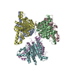
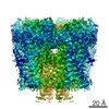
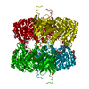
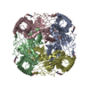
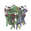
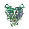
| Title | Cryo-EM structure of polycystic kidney disease-like channel PKD2L1 | |||||||||||||||||||||||||||||||||
 Components Components | Polycystic kidney disease 2-like 1 protein | |||||||||||||||||||||||||||||||||
 Keywords Keywords | MEMBRANE PROTEIN / channel / cryo-EM | |||||||||||||||||||||||||||||||||
| Function / homology |  Function and homology information Function and homology informationdetection of chemical stimulus involved in sensory perception of sour taste / detection of chemical stimulus involved in sensory perception of taste / sensory perception of sour taste / response to water / osmolarity-sensing monoatomic cation channel activity / pH-gated monoatomic ion channel activity / calcium-activated potassium channel activity / cellular response to pH / muscle alpha-actinin binding / detection of mechanical stimulus ...detection of chemical stimulus involved in sensory perception of sour taste / detection of chemical stimulus involved in sensory perception of taste / sensory perception of sour taste / response to water / osmolarity-sensing monoatomic cation channel activity / pH-gated monoatomic ion channel activity / calcium-activated potassium channel activity / cellular response to pH / muscle alpha-actinin binding / detection of mechanical stimulus / cation channel complex / non-motile cilium / calcium-activated cation channel activity / : / ciliary membrane / smoothened signaling pathway / sodium channel activity / alpha-actinin binding / monoatomic cation transmembrane transport / monoatomic cation transport / monoatomic cation channel activity / cellular response to acidic pH / cytoskeletal protein binding / calcium channel complex / potassium ion transmembrane transport / sodium ion transmembrane transport / protein tetramerization / calcium channel activity / actin cytoskeleton / cytoplasmic vesicle / protein homotetramerization / transmembrane transporter binding / signaling receptor complex / calcium ion binding / endoplasmic reticulum / membrane / identical protein binding / plasma membrane / cytosol Similarity search - Function | |||||||||||||||||||||||||||||||||
| Biological species |  | |||||||||||||||||||||||||||||||||
| Method | ELECTRON MICROSCOPY / single particle reconstruction / cryo EM / Resolution: 3.38 Å | |||||||||||||||||||||||||||||||||
 Authors Authors | Zhang, Y.Q. | |||||||||||||||||||||||||||||||||
| Funding support |  China, 1items China, 1items
| |||||||||||||||||||||||||||||||||
 Citation Citation | Journal: Nat Commun / Year: 2018 Title: Cryo-EM structure of the polycystic kidney disease-like channel PKD2L1. Authors: Qiang Su / Feizhuo Hu / Yuxia Liu / Xiaofei Ge / Changlin Mei / Shengqiang Yu / Aiwen Shen / Qiang Zhou / Chuangye Yan / Jianlin Lei / Yanqing Zhang / Xiaodong Liu / Tingliang Wang / Abstract: PKD2L1, also termed TRPP3 from the TRPP subfamily (polycystic TRP channels), is involved in the sour sensation and other pH-dependent processes. PKD2L1 is believed to be a nonselective cation channel ...PKD2L1, also termed TRPP3 from the TRPP subfamily (polycystic TRP channels), is involved in the sour sensation and other pH-dependent processes. PKD2L1 is believed to be a nonselective cation channel that can be regulated by voltage, protons, and calcium. Despite its considerable importance, the molecular mechanisms underlying PKD2L1 regulations are largely unknown. Here, we determine the PKD2L1 atomic structure at 3.38 Å resolution by cryo-electron microscopy, whereby side chains of nearly all residues are assigned. Unlike its ortholog PKD2, the pore helix (PH) and transmembrane segment 6 (S6) of PKD2L1, which are involved in upper and lower-gate opening, adopt an open conformation. Structural comparisons of PKD2L1 with a PKD2-based homologous model indicate that the pore domain dilation is coupled to conformational changes of voltage-sensing domains (VSDs) via a series of π-π interactions, suggesting a potential PKD2L1 gating mechanism. | |||||||||||||||||||||||||||||||||
| History |
|
- Structure visualization
| Movie |
Movie viewer |
|---|---|
| Structure viewer | Molecule: MolmilJmol/JSmol |
- Downloads & links
-Download
| PDBx/mmCIF format | 5z1w.cif.gz | 315.4 KB | Display | PDBx/mmCIF format |
|---|---|---|---|---|
| PDB format | pdb5z1w.ent.gz | 252 KB | Display | PDB format |
| PDBx/mmJSON format | 5z1w.json.gz | Tree view | PDBx/mmJSON format | |
| Others |  Other downloads Other downloads |
-Validation report
| Arichive directory | https://data.pdbj.org/pub/pdb/validation_reports/z1/5z1wftp://data.pdbj.org/pub/pdb/validation_reports/z1/5z1w | HTTPS FTP |
|---|
-Related structure data
| Related structure data |  6877MC M: map data used to model this data C: citing same article ( |
|---|---|
| Similar structure data |

-Links
 PDBj
PDBj
- Assembly
| Deposited unit | 
|
|---|---|
| 1 |
|
-Components
| #1: Protein | Mass: 65600.883 Da / Num. of mol.: 4 Source method: isolated from a genetically manipulated source Source: (gene. exp.)  Homo sapiens (human) / References: UniProt: A2A259 Homo sapiens (human) / References: UniProt: A2A259#2: Sugar | ChemComp-NAG /   Type: D-saccharide, beta linking / Mass: 221.208 Da / Num. of mol.: 12 Type: D-saccharide, beta linking / Mass: 221.208 Da / Num. of mol.: 12Source method: isolated from a genetically manipulated source Formula: C8H15NO6 Has protein modification | Y | |
|---|
-Experimental details
-Experiment
| Experiment | Method: ELECTRON MICROSCOPY |
|---|---|
| EM experiment | Aggregation state: PARTICLE / 3D reconstruction method: single particle reconstruction |
- Sample preparation
| Component | Name: PKD2L1 / Type: COMPLEX / Entity ID: #1 / Source: RECOMBINANT |
|---|---|
| Source (natural) | Organism: |
| Source (recombinant) | Organism: Homo sapiens (human) / Cell: HEK293F |
| Buffer solution | pH: 7.5 |
| Specimen | Embedding applied: NO / Shadowing applied: NO / Staining applied: NO / Vitrification applied: YES |
| Specimen support | Grid material: COPPER / Grid mesh size: 300 divisions/in. / Grid type: Quantifoil R1.2/1.3 |
| Vitrification | Instrument: FEI VITROBOT MARK IV / Cryogen name: ETHANE / Humidity: 100 % |
- Electron microscopy imaging
| Experimental equipment |  Model: Titan Krios / Image courtesy: FEI Company |
|---|---|
| Microscopy | Model: FEI TITAN KRIOS |
| Electron gun | Electron source:  FIELD EMISSION GUN / Accelerating voltage: 300 kV / Illumination mode: FLOOD BEAM FIELD EMISSION GUN / Accelerating voltage: 300 kV / Illumination mode: FLOOD BEAM |
| Electron lens | Mode: BRIGHT FIELD / Cs: 0.001 mm |
| Image recording | Electron dose: 60 e/Å2 / Film or detector model: FEI FALCON II (4k x 4k) |
- Processing
| Software | Name: PHENIX / Version: 1.10.1_2155: / Classification: refinement | ||||||||||||||||||||||||
|---|---|---|---|---|---|---|---|---|---|---|---|---|---|---|---|---|---|---|---|---|---|---|---|---|---|
| EM software | Name: PHENIX / Category: model refinement | ||||||||||||||||||||||||
| CTF correction | Type: NONE | ||||||||||||||||||||||||
| 3D reconstruction | Resolution: 3.38 Å / Resolution method: FSC 0.143 CUT-OFF / Num. of particles: 22296 / Symmetry type: POINT | ||||||||||||||||||||||||
| Refine LS restraints |
|
