 Movie
Movie Controller

[English] 日本語
 Yorodumi
- EMDB-6877: Cryo-EM structure of polycystic kidney disease-like channel PKD2L1 -
+ Open data

- Basic information
| Entry | Database: EMDB / ID: EMD-6877 | |||||||||
|---|---|---|---|---|---|---|---|---|---|---|
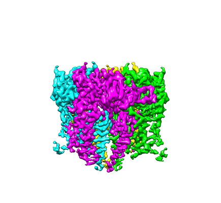
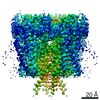
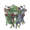
| Title | Cryo-EM structure of polycystic kidney disease-like channel PKD2L1 | |||||||||
 Map data Map data | ||||||||||
 Sample Sample |
| |||||||||
 Keywords Keywords | channel / cryo-EM / MEMBRANE PROTEIN | |||||||||
| Function / homology |  Function and homology information Function and homology informationdetection of chemical stimulus involved in sensory perception of sour taste / detection of chemical stimulus involved in sensory perception of taste / sensory perception of sour taste / response to water / osmolarity-sensing monoatomic cation channel activity / pH-gated monoatomic ion channel activity / calcium-activated potassium channel activity / cellular response to pH / muscle alpha-actinin binding / detection of mechanical stimulus ...detection of chemical stimulus involved in sensory perception of sour taste / detection of chemical stimulus involved in sensory perception of taste / sensory perception of sour taste / response to water / osmolarity-sensing monoatomic cation channel activity / pH-gated monoatomic ion channel activity / calcium-activated potassium channel activity / cellular response to pH / muscle alpha-actinin binding / detection of mechanical stimulus / cation channel complex / non-motile cilium / calcium-activated cation channel activity / : / smoothened signaling pathway / ciliary membrane / sodium channel activity / alpha-actinin binding / monoatomic cation transmembrane transport / monoatomic cation transport / monoatomic cation channel activity / cellular response to acidic pH / cytoskeletal protein binding / calcium channel complex / potassium ion transmembrane transport / sodium ion transmembrane transport / protein tetramerization / calcium channel activity / actin cytoskeleton / cytoplasmic vesicle / protein homotetramerization / transmembrane transporter binding / signaling receptor complex / calcium ion binding / endoplasmic reticulum / membrane / identical protein binding / plasma membrane / cytosol Similarity search - Function | |||||||||
| Biological species |  | |||||||||
| Method | single particle reconstruction / cryo EM / Resolution: 3.38 Å | |||||||||
 Authors Authors | Zhang YQ | |||||||||
 Citation Citation | Journal: Nat Commun / Year: 2018 Title: Cryo-EM structure of the polycystic kidney disease-like channel PKD2L1. Authors: Qiang Su / Feizhuo Hu / Yuxia Liu / Xiaofei Ge / Changlin Mei / Shengqiang Yu / Aiwen Shen / Qiang Zhou / Chuangye Yan / Jianlin Lei / Yanqing Zhang / Xiaodong Liu / Tingliang Wang /  Abstract: PKD2L1, also termed TRPP3 from the TRPP subfamily (polycystic TRP channels), is involved in the sour sensation and other pH-dependent processes. PKD2L1 is believed to be a nonselective cation channel ...PKD2L1, also termed TRPP3 from the TRPP subfamily (polycystic TRP channels), is involved in the sour sensation and other pH-dependent processes. PKD2L1 is believed to be a nonselective cation channel that can be regulated by voltage, protons, and calcium. Despite its considerable importance, the molecular mechanisms underlying PKD2L1 regulations are largely unknown. Here, we determine the PKD2L1 atomic structure at 3.38 Å resolution by cryo-electron microscopy, whereby side chains of nearly all residues are assigned. Unlike its ortholog PKD2, the pore helix (PH) and transmembrane segment 6 (S6) of PKD2L1, which are involved in upper and lower-gate opening, adopt an open conformation. Structural comparisons of PKD2L1 with a PKD2-based homologous model indicate that the pore domain dilation is coupled to conformational changes of voltage-sensing domains (VSDs) via a series of π-π interactions, suggesting a potential PKD2L1 gating mechanism. | |||||||||
| History |
|
- Structure visualization
| Movie |
Movie viewer |
|---|---|
| Structure viewer | EM map: SurfViewMolmilJmol/JSmol |
| Supplemental images |
- Downloads & links
-EMDB archive
| Map data | emd_6877.map.gz | 95.4 MB | EMDB map data format | |
|---|---|---|---|---|
| Header (meta data) | emd-6877-v30.xmlemd-6877.xml | 14.4 KB 14.4 KB | Display Display | EMDB header |
| Images |  emd_6877.png emd_6877.png | 97.8 KB | ||
| Filedesc metadata | emd-6877.cif.gz | 6.1 KB | ||
| Archive directory |  http://ftp.pdbj.org/pub/emdb/structures/EMD-6877ftp://ftp.pdbj.org/pub/emdb/structures/EMD-6877 http://ftp.pdbj.org/pub/emdb/structures/EMD-6877ftp://ftp.pdbj.org/pub/emdb/structures/EMD-6877 | HTTPS FTP |
-Related structure data
| Related structure data |  5z1wMC M: atomic model generated by this map C: citing same article ( |
|---|---|
| Similar structure data |


-Links
| EMDB pages | EMDB (EBI/PDBe) / EMDataResource |
|---|---|
| Related items in Molecule of the Month |

-Map
| File | Download / File: emd_6877.map.gz / Format: CCP4 / Size: 103 MB / Type: IMAGE STORED AS FLOATING POINT NUMBER (4 BYTES) | ||||||||||||||||||||||||||||||||||||||||||||||||||||||||||||
|---|---|---|---|---|---|---|---|---|---|---|---|---|---|---|---|---|---|---|---|---|---|---|---|---|---|---|---|---|---|---|---|---|---|---|---|---|---|---|---|---|---|---|---|---|---|---|---|---|---|---|---|---|---|---|---|---|---|---|---|---|---|
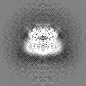
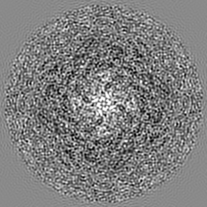
| Projections & slices | Image control
Images are generated by Spider. | ||||||||||||||||||||||||||||||||||||||||||||||||||||||||||||
| Voxel size | X=Y=Z: 0.88 Å | ||||||||||||||||||||||||||||||||||||||||||||||||||||||||||||
| Density |
| ||||||||||||||||||||||||||||||||||||||||||||||||||||||||||||
| Symmetry | Space group: 1 | ||||||||||||||||||||||||||||||||||||||||||||||||||||||||||||
| Details | EMDB XML:
CCP4 map header:
| ||||||||||||||||||||||||||||||||||||||||||||||||||||||||||||
 Z (Sec.)
 Y (Row.)
 X (Col.)


-Supplemental data
- Sample components
-Entire : PKD2L1
| Entire | Name: PKD2L1 |
|---|---|
| Components |
|
-Supramolecule #1: PKD2L1
| Supramolecule | Name: PKD2L1 / type: complex / ID: 1 / Parent: 0 / Macromolecule list: #1 |
|---|---|
| Source (natural) | Organism: |
-Macromolecule #1: Polycystic kidney disease 2-like 1 protein
| Macromolecule | Name: Polycystic kidney disease 2-like 1 protein / type: protein_or_peptide / ID: 1 / Number of copies: 4 / Enantiomer: LEVO |
|---|---|
| Source (natural) | Organism: |
| Molecular weight | Theoretical: 65.600883 KDa |
| Recombinant expression | Organism:  Homo sapiens (human) Homo sapiens (human) |
| Sequence | String: TLVSSCCLHI CRSIRGLWGT TLTENTAENR ELYVKTTLRE LVVYIVFLVD ICLLTYGMTS SSAYYYTKVM SELFLHTPSD SGVSFQTIS SMSDFWDFAQ GPLLDSLYWT KWYNNQSLGR GSHSFIYYEN LLLGAPRLRQ LRVRNDSCVV HEDFREDILN C YDVYSPDK ...String: TLVSSCCLHI CRSIRGLWGT TLTENTAENR ELYVKTTLRE LVVYIVFLVD ICLLTYGMTS SSAYYYTKVM SELFLHTPSD SGVSFQTIS SMSDFWDFAQ GPLLDSLYWT KWYNNQSLGR GSHSFIYYEN LLLGAPRLRQ LRVRNDSCVV HEDFREDILN C YDVYSPDK EDQLPFGPQN GTAWTYHSQN ELGGSSHWGR LTSYSGGGYY LDLPGSRQAS AEALQGLQEG LWLDRGTRVV FI DFSVYNA NINLFCILRL VVEFPATGGT IPSWQIRTVK LIRYVNNWDF FIVGCEVVFC VFIFYYVVEE ILEIHLHRLR YLS SVWNIL DLVVILLSIV AVGFHIFRTL EVNRLMGKLL QQPDTYADFE FLAFWQTQYN NMNAVNLFFA WIKIFKYISF NKTM TQLSS TLARCAKDIL GFAIMFFIVF FAYAQLGYLL FGTQVENFST FVKCIFTQFR IILGDFDYNA IDNANRILGP VYFVT YVFF VFFVLLNMFL AIINDTYSEV KEELAGQKDQ LQLSDFLKQS YNKTLLRLRL RKERVSDVQK VLKGGEPEIQ FEDFTS TLR ELG UniProtKB: Polycystin-2-like protein 1 |
-Macromolecule #2: 2-acetamido-2-deoxy-beta-D-glucopyranose
| Macromolecule | Name: 2-acetamido-2-deoxy-beta-D-glucopyranose / type: ligand / ID: 2 / Number of copies: 12 / Formula: NAG |
|---|---|
| Molecular weight | Theoretical: 221.208 Da |
| Chemical component information |  ChemComp-NAG: |
-Experimental details
-Structure determination
| Method | cryo EM |
|---|---|
 Processing Processing | single particle reconstruction |
| Aggregation state | particle |
-Sample preparation
| Buffer | pH: 7.5 |
|---|---|
| Grid | Model: Quantifoil R1.2/1.3 / Material: COPPER / Mesh: 300 / Pretreatment - Type: GLOW DISCHARGE / Pretreatment - Time: 30 sec. |
| Vitrification | Cryogen name: ETHANE / Chamber humidity: 100 % / Instrument: FEI VITROBOT MARK IV |
- Electron microscopy
| Microscope | FEI TITAN KRIOS |
|---|---|
| Image recording | Film or detector model: FEI FALCON II (4k x 4k) / Average electron dose: 60.0 e/Å2 |
| Electron beam | Acceleration voltage: 300 kV / Electron source:  FIELD EMISSION GUN FIELD EMISSION GUN |
| Electron optics | Illumination mode: FLOOD BEAM / Imaging mode: BRIGHT FIELD / Cs: 0.001 mm |
| Experimental equipment |  Model: Titan Krios / Image courtesy: FEI Company |
