National Institutes of Health/National Institute of Diabetes and Digestive and Kidney Disease (NIH/NIDDK)
R01 DK110575
United States
Citation
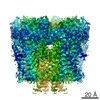
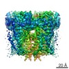
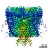
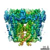
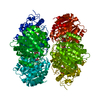
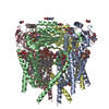
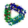
Journal: Proc Natl Acad Sci U S A / Year: 2020 Title: Molecular dysregulation of ciliary polycystin-2 channels caused by variants in the TOP domain. Authors: Thuy N Vien / Jinliang Wang / Leo C T Ng / Erhu Cao / Paul G DeCaen / Abstract: Genetic variants in which encodes for the polycystin-2 ion channel are responsible for many clinical cases of autosomal dominant polycystic kidney disease (ADPKD). Despite our strong understanding ...Genetic variants in which encodes for the polycystin-2 ion channel are responsible for many clinical cases of autosomal dominant polycystic kidney disease (ADPKD). Despite our strong understanding of the genetic basis of ADPKD, we do not know how most variants impact channel function. Polycystin-2 is found in organelle membranes, including the primary cilium-an antennae-like structure on the luminal side of the collecting duct. In this study, we focus on the structural and mechanistic regulation of polycystin-2 by its TOP domain-a site with unknown function that is commonly altered by missense variants. We use direct cilia electrophysiology, cryogenic electron microscopy, and superresolution imaging to determine that variants of the TOP domain finger 1 motif destabilizes the channel structure and impairs channel opening without altering cilia localization and channel assembly. Our findings support the channelopathy classification of variants associated with ADPKD, where polycystin-2 channel dysregulation in the primary cilia may contribute to cystogenesis.
History
Deposition
Mar 26, 2020
-
Header (metadata) release
Apr 22, 2020
-
Map release
Apr 22, 2020
-
Update
May 21, 2025
-
Current status
May 21, 2025
Processing site: RCSB / Status: Released
-
Structure visualization
Movie
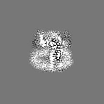
Surface view with section colored by density value
In the structure databanks used in Yorodumi, some data are registered as the other names, "COVID-19 virus" and "2019-nCoV". Here are the details of the virus and the list of structure data.
Jan 31, 2019. EMDB accession codes are about to change! (news from PDBe EMDB page)
EMDB accession codes are about to change! (news from PDBe EMDB page)
The allocation of 4 digits for EMDB accession codes will soon come to an end. Whilst these codes will remain in use, new EMDB accession codes will include an additional digit and will expand incrementally as the available range of codes is exhausted. The current 4-digit format prefixed with “EMD-” (i.e. EMD-XXXX) will advance to a 5-digit format (i.e. EMD-XXXXX), and so on. It is currently estimated that the 4-digit codes will be depleted around Spring 2019, at which point the 5-digit format will come into force.
The EM Navigator/Yorodumi systems omit the EMD- prefix.
Related info.:Q: What is EMD? / ID/Accession-code notation in Yorodumi/EM Navigator
Yorodumi is a browser for structure data from EMDB, PDB, SASBDB, etc.
This page is also the successor to EM Navigator detail page, and also detail information page/front-end page for Omokage search.
The word "yorodu" (or yorozu) is an old Japanese word meaning "ten thousand". "mi" (miru) is to see.
Related info.:EMDB / PDB / SASBDB / Comparison of 3 databanks / Yorodumi Search / Aug 31, 2016. New EM Navigator & Yorodumi / Yorodumi Papers / Jmol/JSmol / Function and homology information / Changes in new EM Navigator and Yorodumi
 Movie
Movie Controller


 Open data
 Basic information
 Map data
 Sample
 Keywords
 Function and homology information
 Homo sapiens (human)
 Authors
 United States, 1 items
  Citation
 Structure visualization
 Downloads & links
 emd_21586.png
 http://ftp.pdbj.org/pub/emdb/structures/EMD-21586


 Z (Sec.)
 Y (Row.)
 X (Col.)


 Sample components
 Processing
 Electron microscopy
 FIELD EMISSION GUN
