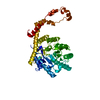
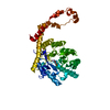
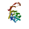
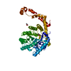
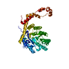
Entry Database : PDB / ID : 5avnTitle The 1.03 angstrom structure (P212121) of glucose isomerase crystallized in high-strength agarose hydrogel Xylose isomerase Keywords / / Function / homology Function Domain/homology Component
/ / / / / / / / / / / / / / Biological species Streptomyces rubiginosus (bacteria)Method / / / Resolution : 1.03 Å Authors Sugiyama, S. / Shimizu, N. / Maruyama, N. / Sazaki, G. / Adachi, H. / Takano, K. / Murakami, S. / Inoue, T. / Mori, Y. / Matsumura, H. Funding support Organization Grant number Country JSPS KAKENHI 23860028 JSPS KAKENHI 25650051 JSPS KAKENHI 25286051
Journal : J.Am.Chem.Soc. / Year : 2012Title : Growth of protein crystals in hydrogels prevents osmotic shockAuthors : Sugiyama, S. / Maruyama, M. / Sazaki, G. / Hirose, M. / Adachi, H. / Takano, K. / Murakami, S. / Inoue, T. / Mori, Y. / Matsumura, H. History Deposition Jun 23, 2015 Deposition site / Processing site Revision 1.0 Jul 8, 2015 Provider / Type Revision 1.1 Feb 26, 2020 Group Advisory / Data collection ... Advisory / Data collection / Derived calculations / Source and taxonomy Category diffrn_source / entity_src_gen ... diffrn_source / entity_src_gen / pdbx_prerelease_seq / pdbx_struct_assembly / pdbx_struct_assembly_gen / pdbx_struct_assembly_prop / pdbx_struct_oper_list / pdbx_validate_close_contact / pdbx_validate_symm_contact Item _diffrn_source.pdbx_synchrotron_site / _entity_src_gen.pdbx_alt_source_flag ... _diffrn_source.pdbx_synchrotron_site / _entity_src_gen.pdbx_alt_source_flag / _pdbx_struct_assembly.oligomeric_details / _pdbx_struct_assembly_gen.asym_id_list / _pdbx_struct_assembly_prop.type / _pdbx_struct_assembly_prop.value / _pdbx_struct_oper_list.symmetry_operation Revision 1.2 Nov 8, 2023 Group Data collection / Database references ... Data collection / Database references / Derived calculations / Refinement description Category chem_comp_atom / chem_comp_bond ... chem_comp_atom / chem_comp_bond / database_2 / pdbx_initial_refinement_model / struct_conn Item _database_2.pdbx_DOI / _database_2.pdbx_database_accession ... _database_2.pdbx_DOI / _database_2.pdbx_database_accession / _struct_conn.pdbx_dist_value / _struct_conn.ptnr1_auth_asym_id / _struct_conn.ptnr1_auth_comp_id / _struct_conn.ptnr1_auth_seq_id / _struct_conn.ptnr1_label_asym_id / _struct_conn.ptnr1_label_atom_id / _struct_conn.ptnr1_label_comp_id / _struct_conn.ptnr1_label_seq_id / _struct_conn.ptnr2_auth_asym_id / _struct_conn.ptnr2_auth_comp_id / _struct_conn.ptnr2_auth_seq_id / _struct_conn.ptnr2_label_asym_id / _struct_conn.ptnr2_label_atom_id / _struct_conn.ptnr2_label_comp_id
Show all Show less
 Movie
Movie Controller

 Yorodumi
 Open data
 Basic information
 Components
 Keywords
 Function and homology information
 Streptomyces rubiginosus (bacteria)
 X-RAY DIFFRACTION /
  Authors
 Japan, 3items
  Citation
 Structure visualization
 Downloads & links
 Other downloads
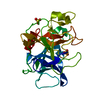


 PDBj
PDBj
 Assembly


 Mass: 54.938 Da / Num. of mol.: 2 / Source method: obtained synthetically / Formula: Mn
 Mass: 40.078 Da / Num. of mol.: 2 / Source method: obtained synthetically / Formula: Ca
 Mass: 96.063 Da / Num. of mol.: 4 / Source method: obtained synthetically / Formula: SO4
 Mass: 18.015 Da / Num. of mol.: 1058 / Source method: isolated from a natural source / Formula: H2O
 Sample preparation
 Processing