 Movie
Movie Controller

[English] 日本語
 Yorodumi
- PDB-6ll2: Crystal structure of glucose isomerase by fixed-target serial fem... -
+ Open data

- Basic information
| Entry | Database: PDB / ID: 6ll2 | ||||||
|---|---|---|---|---|---|---|---|
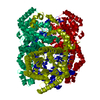
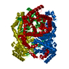
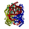
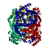
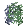
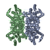
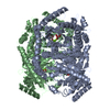
| Title | Crystal structure of glucose isomerase by fixed-target serial femtosecond crystallography | ||||||
 Components Components | Xylose isomerase | ||||||
 Keywords Keywords | ISOMERASE / glucose isomerase / serial crystallography / sfx | ||||||
| Function / homology |  Function and homology information Function and homology informationxylose isomerase / xylose isomerase activity / D-xylose metabolic process / magnesium ion binding / identical protein binding / cytoplasm Similarity search - Function | ||||||
| Biological species |  Streptomyces rubiginosus (bacteria) Streptomyces rubiginosus (bacteria) | ||||||
| Method |  X-RAY DIFFRACTION / FREE ELECTRON LASER / MOLECULAR REPLACEMENT / molecular replacement / Resolution: 1.75 Å X-RAY DIFFRACTION / FREE ELECTRON LASER / MOLECULAR REPLACEMENT / molecular replacement / Resolution: 1.75 Å | ||||||
 Authors Authors | Nam, K.H. | ||||||
| Funding support |  Korea, Republic Of, 1items Korea, Republic Of, 1items
| ||||||
 Citation Citation | Journal: To Be Published Title: Crystal structure of glucose isomerase by fixed-target serial femtosecond crystallography Authors: Nam, K.H. | ||||||
| History |
|
- Structure visualization
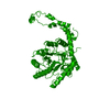
| Structure viewer | Molecule: MolmilJmol/JSmol |
|---|
- Downloads & links
-Download
| PDBx/mmCIF format | 6ll2.cif.gz | 100.3 KB | Display | PDBx/mmCIF format |
|---|---|---|---|---|
| PDB format | pdb6ll2.ent.gz | 74.1 KB | Display | PDB format |
| PDBx/mmJSON format | 6ll2.json.gz | Tree view | PDBx/mmJSON format | |
| Others |  Other downloads Other downloads |
-Validation report
| Arichive directory | https://data.pdbj.org/pub/pdb/validation_reports/ll/6ll2ftp://data.pdbj.org/pub/pdb/validation_reports/ll/6ll2 | HTTPS FTP |
|---|
-Related structure data
| Related structure data | 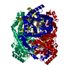 5zyeS S: Starting model for refinement |
|---|---|
| Similar structure data |

-Links
 PDBj
PDBj
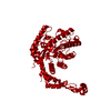
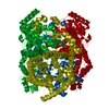
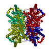
- Assembly
| Deposited unit | 
| |||||||||||||||
|---|---|---|---|---|---|---|---|---|---|---|---|---|---|---|---|---|
| 1 | 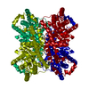
| |||||||||||||||
| Unit cell |
| |||||||||||||||
| Components on special symmetry positions |
|
-Components
| #1: Protein | Mass: 43283.297 Da / Num. of mol.: 1 / Source method: isolated from a natural source / Source: (natural) Streptomyces rubiginosus (bacteria) / References: UniProt: P24300, xylose isomerase | ||||
|---|---|---|---|---|---|
| #2: Chemical |   Mass: 24.305 Da / Num. of mol.: 2 / Source method: obtained synthetically / Formula: Mg Mass: 24.305 Da / Num. of mol.: 2 / Source method: obtained synthetically / Formula: Mg#3: Water | ChemComp-HOH / |  Mass: 18.015 Da / Num. of mol.: 387 / Source method: isolated from a natural source / Formula: H2O Mass: 18.015 Da / Num. of mol.: 387 / Source method: isolated from a natural source / Formula: H2OHas ligand of interest | N | |
-Experimental details
-Experiment
| Experiment | Method: X-RAY DIFFRACTION / Number of used crystals: 1 |
|---|
- Sample preparation
| Crystal | Density Matthews: 2.67 Å3/Da / Density % sol: 53.93 % |
|---|---|
| Crystal grow | Temperature: 293.5 K / Method: batch mode / pH: 7 / Details: Tris-HCl, Ammonium sulfate, Magnesium sulfate |
-Data collection
| Diffraction | Mean temperature: 297 K / Serial crystal experiment: Y |
|---|---|
| Diffraction source | Source: FREE ELECTRON LASER / Site: PAL-XFEL / Beamline: NCI / Wavelength: 1.2782 Å |
| Detector | Type: RAYONIX MX225-HS / Detector: CCD / Date: Nov 10, 2019 / Details: Kirkpatrick-Baez mirror |
| Radiation | Protocol: SINGLE WAVELENGTH / Monochromatic (M) / Laue (L): M / Scattering type: x-ray |
| Radiation wavelength | Wavelength: 1.2782 Å / Relative weight: 1 |
| Reflection | Resolution: 1.75→70.9 Å / Num. obs: 47004 / % possible obs: 100 % / Redundancy: 346.2 % / CC1/2: 0.934777 / CC star: 0.9397 / Net I/σ(I): 5.2 |
| Reflection shell | Resolution: 1.75→1.81 Å / Redundancy: 124.5 % / Mean I/σ(I) obs: 3 / Num. unique obs: 4650 / CC1/2: 0.7908 / CC star: 0.9397 / % possible all: 100 |
| Serial crystallography measurement | XFEL pulse repetition rate: 30 Hz |
| Serial crystallography sample delivery | Method: fixed target |
| Serial crystallography sample delivery fixed target | Sample dehydration prevention: polyimide sealing / Sample holding: viscous medium / Support base: x-y translation stage |
| Serial crystallography data reduction | Frame hits: 33438 / Frames indexed: 22412 / Frames total: 55307 |
-Phasing
| Phasing | Method: molecular replacement |
|---|
- Processing
| Software |
| |||||||||||||||||||||||||||||||||||||||||||||||||||||||
|---|---|---|---|---|---|---|---|---|---|---|---|---|---|---|---|---|---|---|---|---|---|---|---|---|---|---|---|---|---|---|---|---|---|---|---|---|---|---|---|---|---|---|---|---|---|---|---|---|---|---|---|---|---|---|---|---|
| Refinement | Method to determine structure: MOLECULAR REPLACEMENT Starting model: 5ZYE Resolution: 1.75→70.623 Å / SU ML: 0.16 / Cross valid method: THROUGHOUT / σ(F): 1.33 / Phase error: 17.51
| |||||||||||||||||||||||||||||||||||||||||||||||||||||||
| Solvent computation | Shrinkage radii: 0.9 Å / VDW probe radii: 1.11 Å | |||||||||||||||||||||||||||||||||||||||||||||||||||||||
| Displacement parameters | Biso max: 62.8 Å2 / Biso mean: 13.1837 Å2 / Biso min: 1.17 Å2 | |||||||||||||||||||||||||||||||||||||||||||||||||||||||
| Refinement step | Cycle: final / Resolution: 1.75→70.623 Å
| |||||||||||||||||||||||||||||||||||||||||||||||||||||||
| LS refinement shell | Refine-ID: X-RAY DIFFRACTION / Rfactor Rfree error: 0 / % reflection obs: 100 %
|
