Protocol: SINGLE WAVELENGTH / Monochromatic (M) / Laue (L): M / Scattering type: x-ray
Radiation wavelength
Wavelength: 0.7 Å / Relative weight: 1
Reflection
Resolution: 0.9→50 Å / Num. obs: 325304 / % possible obs: 95.1 % / Redundancy: 3 % / Rmerge(I) obs: 0.073 / Net I/σ(I): 11.6
Reflection shell
Resolution: 0.9→0.92 Å / Redundancy: 2.2 % / Rmerge(I) obs: 0.354 / Mean I/σ(I) obs: 2.38 / % possible all: 94.3
-
Processing
Software
Name
Version
Classification
REFMAC
5.8.0049
refinement
DENZO
datareduction
HKL-2000
datascaling
MOLREP
phasing
Refinement
Resolution: 0.9→50 Å / Cor.coef. Fo:Fc: 0.981 / Cor.coef. Fo:Fc free: 0.979 / SU B: 0.344 / SU ML: 0.009 / Cross valid method: THROUGHOUT / ESU R: 0.013 / ESU R Free: 0.014 / Stereochemistry target values: MAXIMUM LIKELIHOOD / Details: HYDROGENS HAVE BEEN ADDED IN THE RIDING POSITIONS
Rfactor
Num. reflection
% reflection
Selection details
Rfree
0.12735
16000
5 %
RANDOM
Rwork
0.11628
-
-
-
obs
0.11683
300000
95.11 %
-
Solvent computation
Ion probe radii: 0.8 Å / Shrinkage radii: 0.8 Å / VDW probe radii: 1.2 Å / Solvent model: MASK
 Movie
Movie Controller

 Yorodumi
 Open data
 Basic information
 Components
 Keywords
 Function and homology information
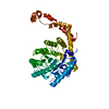
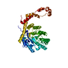
 Streptomyces rubiginosus (bacteria)
 X-RAY DIFFRACTION /
  Authors
 Japan, 3items
  Citation
 Structure visualization
 Downloads & links
 Other downloads


 PDBj
PDBj
 Assembly
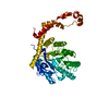
 Mass: 18.015 Da / Num. of mol.: 515 / Source method: isolated from a natural source / Formula: H2O
 Sample preparation
 Processing