[English] 日本語
Yorodumi
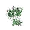
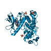
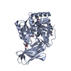
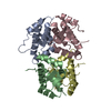
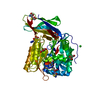
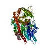
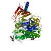
- PDB-5ak8: Structure of C351A mutant of Porphyromonas gingivalis peptidylarg... -

+
Open data


ID or keywords:

Loading...

-
Basic information

Entry
Database: PDB / ID: 5ak8
TitleStructure of C351A mutant of Porphyromonas gingivalis peptidylarginine deiminase
ComponentsPEPTIDYLARGININE DEIMINASE
KeywordsHYDROLASE / PPAD
Function / homology
Function and homology information


Hydrolases; Acting on carbon-nitrogen bonds, other than peptide bonds; In linear amidines / agmatine deiminase activity / putrescine biosynthetic process / protein-arginine deiminase activity / extracellular region
Similarity search - Function
Peptidyl-arginine deiminase, Porphyromonas-type / Porphyromonas-type peptidyl-arginine deiminase / L-arginine/glycine Amidinotransferase; Chain A / 5-stranded Propeller / L-arginine/glycine Amidinotransferase; Chain A / Alpha Beta
Similarity search - Domain/homology
ALANINE / ARGININE / Peptidylarginine deiminase
Similarity search - Component
Biological speciesPORPHYROMONAS GINGIVALIS (bacteria)
MethodX-RAY DIFFRACTION / SYNCHROTRON / MOLECULAR REPLACEMENT / Resolution: 1.48 Å
AuthorsKopec, J. / Montgomery, A. / Shrestha, L. / Kiyani, W. / Nowak, R. / Burgess-Brown, N. / Venables, P.J. / Yue, W.W.
CitationJournal: Ann.Rheum.Dis. / Year: 2016
Title: Crystal Structure of Porphyromonas Gingivalis Peptidylarginine Deiminase: Implications for Autoimmunity in Rheumatoid Arthritis.
Authors: Montgomery, A.B. / Kopec, J. / Shrestha, L. / Thezenas, M.L. / Burgess-Brown, N.A. / Fischer, R. / Yue, W.W. / Venables, P.J.
History
DepositionMar 2, 2015Deposition site: PDBE / Processing site: PDBE
Revision 1.0Jul 22, 2015Provider: repository / Type: Initial release
Revision 1.1Aug 5, 2015Group: Database references
Revision 1.2Aug 19, 2015Group: Structure summary
Revision 1.3May 25, 2016Group: Database references
Revision 1.4Jun 20, 2018Group: Advisory / Data collection / Derived calculations
Category: pdbx_struct_conn_angle / pdbx_unobs_or_zero_occ_atoms ...pdbx_struct_conn_angle / pdbx_unobs_or_zero_occ_atoms / pdbx_validate_close_contact / struct_conn / struct_conn_type
Item: _pdbx_struct_conn_angle.ptnr1_auth_comp_id / _pdbx_struct_conn_angle.ptnr1_auth_seq_id ..._pdbx_struct_conn_angle.ptnr1_auth_comp_id / _pdbx_struct_conn_angle.ptnr1_auth_seq_id / _pdbx_struct_conn_angle.ptnr1_label_asym_id / _pdbx_struct_conn_angle.ptnr1_label_atom_id / _pdbx_struct_conn_angle.ptnr1_label_comp_id / _pdbx_struct_conn_angle.ptnr1_label_seq_id / _pdbx_struct_conn_angle.ptnr3_auth_comp_id / _pdbx_struct_conn_angle.ptnr3_auth_seq_id / _pdbx_struct_conn_angle.ptnr3_label_asym_id / _pdbx_struct_conn_angle.ptnr3_label_atom_id / _pdbx_struct_conn_angle.ptnr3_label_comp_id / _pdbx_struct_conn_angle.ptnr3_label_seq_id / _pdbx_struct_conn_angle.value
Revision 1.5May 8, 2024Group: Data collection / Database references ...Data collection / Database references / Derived calculations / Other
Category: chem_comp_atom / chem_comp_bond ...chem_comp_atom / chem_comp_bond / database_2 / pdbx_database_status / pdbx_struct_conn_angle / struct_conn / struct_site
Item: _database_2.pdbx_DOI / _database_2.pdbx_database_accession ..._database_2.pdbx_DOI / _database_2.pdbx_database_accession / _pdbx_database_status.status_code_sf / _pdbx_struct_conn_angle.ptnr1_auth_seq_id / _pdbx_struct_conn_angle.ptnr3_auth_seq_id / _pdbx_struct_conn_angle.value / _struct_conn.pdbx_dist_value / _struct_conn.ptnr2_auth_seq_id / _struct_site.pdbx_auth_asym_id / _struct_site.pdbx_auth_comp_id / _struct_site.pdbx_auth_seq_id

-
Structure visualization

Structure viewerMolecule:
MolmilJmol/JSmol

Downloads & links

-
Assembly

Deposited unit
A: PEPTIDYLARGININE DEIMINASE
hetero molecules


Theoretical massNumber of molelcules
Total (without water)50,31122
Polymers48,9071
Non-polymers1,40521
Water8,323462
1


  • Idetical with deposited unit
  • defined by author&software
TypeNameSymmetry operationNumber
identity operation1_555x,y,z1
MethodPISA
Unit cell
Length a, b, c (Å)101.790, 84.090, 55.460
Angle α, β, γ (deg.)90.00, 92.64, 90.00
Int Tables number5
Space group name H-MC121

-
Components

-
Protein , 1 types, 1 molecules A

#1: Protein PEPTIDYLARGININE DEIMINASE / PPAD


Mass: 48906.645 Da / Num. of mol.: 1 / Fragment: RESIDUES 49-484 / Mutation: YES
Source method: isolated from a genetically manipulated source
Source: (gene. exp.) PORPHYROMONAS GINGIVALIS (bacteria) / Plasmid: PNIC28-BSA / Production host: ESCHERICHIA COLI (E. coli) / Strain (production host): BL21(DE3) / Variant (production host): ROSETTA
References: UniProt: Q9RQJ2, Hydrolases; Acting on carbon-nitrogen bonds, other than peptide bonds; In linear amidines

-
Non-polymers , 5 types, 483 molecules

#2: Chemical ChemComp-ALA / ALANINE


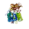
Type: L-peptide linking / Mass: 89.093 Da / Num. of mol.: 1 / Source method: obtained synthetically / Formula: C3H7NO2
#3: Chemical ChemComp-ARG / ARGININE


Type: L-peptide linking / Mass: 175.209 Da / Num. of mol.: 1 / Source method: obtained synthetically / Formula: C6H15N4O2
#4: Chemical ChemComp-NA / SODIUM ION


Mass: 22.990 Da / Num. of mol.: 1 / Source method: obtained synthetically / Formula: Na
#5: Chemical
ChemComp-EDO / 1,2-ETHANEDIOL / ETHYLENE GLYCOL


Mass: 62.068 Da / Num. of mol.: 18 / Source method: obtained synthetically / Formula: C2H6O2
#6: Water ChemComp-HOH / water


Mass: 18.015 Da / Num. of mol.: 462 / Source method: isolated from a natural source / Formula: H2O

-
Details

Sequence detailsC351A - CATALYTIC MUTANT

-
Experimental details

-
Experiment

ExperimentMethod: X-RAY DIFFRACTION / Number of used crystals: 1

-
Sample preparation

CrystalDensity Matthews: 2.43 Å3/Da / Density % sol: 49.34 % / Description: NONE
Crystal growDetails: 0.2M AMMONIUM SULFATE, 30% PEG5000MME, 0.1M MES PH 6.5

-
Data collection

DiffractionMean temperature: 100 K
Diffraction sourceSource: SYNCHROTRON / Site: Diamond / Beamline: I03 / Wavelength: 0.97625
DetectorType: DECTRIS PILATUS 6M / Detector: PIXEL / Date: Apr 25, 2014
RadiationProtocol: SINGLE WAVELENGTH / Monochromatic (M) / Laue (L): M / Scattering type: x-ray
Radiation wavelengthWavelength: 0.97625 Å / Relative weight: 1
ReflectionResolution: 1.48→64.8 Å / Num. obs: 73831 / % possible obs: 95.3 % / Observed criterion σ(I): 1 / Redundancy: 3.4 % / Biso Wilson estimate: 15.2 Å2 / Rmerge(I) obs: 0.06 / Net I/σ(I): 10.4
Reflection shellResolution: 1.48→1.52 Å / Redundancy: 3.6 % / Rmerge(I) obs: 0.54 / Mean I/σ(I) obs: 2.1 / % possible all: 93.9

-
Processing

Software
NameVersionClassification
REFMAC5.8.0107refinement
PHASERphasing
RefinementMethod to determine structure: MOLECULAR REPLACEMENT / Resolution: 1.48→64.8 Å / Cor.coef. Fo:Fc: 0.982 / Cor.coef. Fo:Fc free: 0.973 / SU B: 3.064 / SU ML: 0.049 / Cross valid method: THROUGHOUT / ESU R: 0.064 / ESU R Free: 0.059 / Stereochemistry target values: MAXIMUM LIKELIHOOD
Details: HYDROGENS HAVE BEEN ADDED IN THE RIDING POSITIONS. U VALUES REFINED INDIVIDUALLY
RfactorNum. reflection% reflectionSelection details
Rfree0.16444 3636 4.9 %RANDOM
Rwork0.12737 ---
obs0.12921 70193 95.13 %-
Solvent computationIon probe radii: 0.8 Å / Shrinkage radii: 0.8 Å / VDW probe radii: 1.2 Å / Solvent model: MASK
Displacement parametersBiso mean: 19.703 Å2
Baniso -1Baniso -2Baniso -3
1-0.69 Å2-0 Å2-1.44 Å2
2---0.44 Å20 Å2
3----0.12 Å2
Refinement stepCycle: LAST / Resolution: 1.48→64.8 Å
ProteinNucleic acidLigandSolventTotal
Num. atoms3288 0 90 462 3840
Refine LS restraints
Refine-IDTypeDev idealDev ideal targetNumber
X-RAY DIFFRACTIONr_bond_refined_d0.0130.0193480
X-RAY DIFFRACTIONr_bond_other_d0.0030.023196
X-RAY DIFFRACTIONr_angle_refined_deg1.581.9424705
X-RAY DIFFRACTIONr_angle_other_deg0.98737354
X-RAY DIFFRACTIONr_dihedral_angle_1_deg6.0775431
X-RAY DIFFRACTIONr_dihedral_angle_2_deg34.44224.819166
X-RAY DIFFRACTIONr_dihedral_angle_3_deg11.00715533
X-RAY DIFFRACTIONr_dihedral_angle_4_deg11.1531513
X-RAY DIFFRACTIONr_chiral_restr0.1020.2499
X-RAY DIFFRACTIONr_gen_planes_refined0.0080.0213971
X-RAY DIFFRACTIONr_gen_planes_other0.0020.02810
X-RAY DIFFRACTIONr_nbd_refined
X-RAY DIFFRACTIONr_nbd_other
X-RAY DIFFRACTIONr_nbtor_refined
X-RAY DIFFRACTIONr_nbtor_other
X-RAY DIFFRACTIONr_xyhbond_nbd_refined
X-RAY DIFFRACTIONr_xyhbond_nbd_other
X-RAY DIFFRACTIONr_metal_ion_refined
X-RAY DIFFRACTIONr_metal_ion_other
X-RAY DIFFRACTIONr_symmetry_vdw_refined
X-RAY DIFFRACTIONr_symmetry_vdw_other
X-RAY DIFFRACTIONr_symmetry_hbond_refined
X-RAY DIFFRACTIONr_symmetry_hbond_other
X-RAY DIFFRACTIONr_symmetry_metal_ion_refined
X-RAY DIFFRACTIONr_symmetry_metal_ion_other
X-RAY DIFFRACTIONr_mcbond_it2.6431.7661697
X-RAY DIFFRACTIONr_mcbond_other2.6441.7661696
X-RAY DIFFRACTIONr_mcangle_it3.3122.6632122
X-RAY DIFFRACTIONr_mcangle_other
X-RAY DIFFRACTIONr_scbond_it3.2061.9481783
X-RAY DIFFRACTIONr_scbond_other
X-RAY DIFFRACTIONr_scangle_it
X-RAY DIFFRACTIONr_scangle_other
X-RAY DIFFRACTIONr_long_range_B_refined
X-RAY DIFFRACTIONr_long_range_B_other
X-RAY DIFFRACTIONr_rigid_bond_restr2.47636676
X-RAY DIFFRACTIONr_sphericity_free41.3685156
X-RAY DIFFRACTIONr_sphericity_bonded17.76856909
LS refinement shellResolution: 1.48→1.518 Å / Total num. of bins used: 20
RfactorNum. reflection% reflection
Rfree0.265 242 -
Rwork0.209 5083 -
obs--93.8 %

+
About Yorodumi

-
News

-
Feb 9, 2022. New format data for meta-information of EMDB entries

New format data for meta-information of EMDB entries

  • Version 3 of the EMDB header file is now the official format.
  • The previous official version 1.9 will be removed from the archive.

Related info.:EMDB header

External links:wwPDB to switch to version 3 of the EMDB data model

-
Aug 12, 2020. Covid-19 info

Covid-19 info

URL: https://pdbjlvh1.pdbj.org/emnavi/covid19.php

New page: Covid-19 featured information page in EM Navigator.

Related info.:Covid-19 info / Mar 5, 2020. Novel coronavirus structure data

+
Mar 5, 2020. Novel coronavirus structure data

Novel coronavirus structure data

Related info.:Yorodumi Speices / Aug 12, 2020. Covid-19 info

External links:COVID-19 featured content - PDBj / Molecule of the Month (242):Coronavirus Proteases

+
Jan 31, 2019. EMDB accession codes are about to change! (news from PDBe EMDB page)

EMDB accession codes are about to change! (news from PDBe EMDB page)

  • The allocation of 4 digits for EMDB accession codes will soon come to an end. Whilst these codes will remain in use, new EMDB accession codes will include an additional digit and will expand incrementally as the available range of codes is exhausted. The current 4-digit format prefixed with “EMD-” (i.e. EMD-XXXX) will advance to a 5-digit format (i.e. EMD-XXXXX), and so on. It is currently estimated that the 4-digit codes will be depleted around Spring 2019, at which point the 5-digit format will come into force.
  • The EM Navigator/Yorodumi systems omit the EMD- prefix.

Related info.:Q: What is EMD? / ID/Accession-code notation in Yorodumi/EM Navigator

External links:EMDB Accession Codes are Changing Soon! / Contact to PDBj

+
Jul 12, 2017. Major update of PDB

Major update of PDB

  • wwPDB released updated PDB data conforming to the new PDBx/mmCIF dictionary.
  • This is a major update changing the version number from 4 to 5, and with Remediation, in which all the entries are updated.
  • In this update, many items about electron microscopy experimental information are reorganized (e.g. em_software).
  • Now, EM Navigator and Yorodumi are based on the updated data.

External links:wwPDB Remediation / Enriched Model Files Conforming to OneDep Data Standards Now Available in the PDB FTP Archive

-
Yorodumi

Thousand views of thousand structures

  • Yorodumi is a browser for structure data from EMDB, PDB, SASBDB, etc.
  • This page is also the successor to EM Navigator detail page, and also detail information page/front-end page for Omokage search.
  • The word "yorodu" (or yorozu) is an old Japanese word meaning "ten thousand". "mi" (miru) is to see.

Related info.:EMDB / PDB / SASBDB / Comparison of 3 databanks / Yorodumi Search / Aug 31, 2016. New EM Navigator & Yorodumi / Yorodumi Papers / Jmol/JSmol / Function and homology information / Changes in new EM Navigator and Yorodumi

Read more