 Movie
Movie Controller

[English] 日本語
 Yorodumi
- PDB-1n2l: Crystal structure of a covalent intermediate of endogenous human ... -
+ Open data

- Basic information
| Entry | Database: PDB / ID: 1n2l | |||||||||
|---|---|---|---|---|---|---|---|---|---|---|
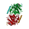
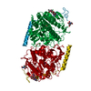
| Title | Crystal structure of a covalent intermediate of endogenous human arylsulfatase A | |||||||||
 Components Components | ARYLSULFATASE A | |||||||||
 Keywords Keywords | HYDROLASE / PHOSPHATE ESTER HYDROLYSIS / LYSOSOMAL ENZYME / MODIFIED FORMYLGLYCINE / INHIBITION / METAL ION | |||||||||
| Function / homology |  Function and homology information Function and homology informationcerebroside-sulfatase / cerebroside-sulfatase activity / The activation of arylsulfatases / sulfuric ester hydrolase activity / arylsulfatase activity / Glycosphingolipid catabolism / lysosomal lumen / lipid metabolic process / azurophil granule lumen / lysosome ...cerebroside-sulfatase / cerebroside-sulfatase activity / The activation of arylsulfatases / sulfuric ester hydrolase activity / arylsulfatase activity / Glycosphingolipid catabolism / lysosomal lumen / lipid metabolic process / azurophil granule lumen / lysosome / endoplasmic reticulum lumen / calcium ion binding / Neutrophil degranulation / extracellular exosome / extracellular region Similarity search - Function | |||||||||
| Biological species |  Homo sapiens (human) Homo sapiens (human) | |||||||||
| Method |  X-RAY DIFFRACTION / MOLECULAR REPLACEMENT / Resolution: 3.2 Å X-RAY DIFFRACTION / MOLECULAR REPLACEMENT / Resolution: 3.2 Å | |||||||||
 Authors Authors | Chruszcz, M. / Laidler, P. / Monkiewicz, M. / Ortlund, E. / Lebioda, L. / Lewinski, K. | |||||||||
 Citation Citation | Journal: J.Inorg.Biochem. / Year: 2003 Title: Crystal structure of a covalent intermediate of endogenous human arylsulfatase A. Authors: Chruszcz, M. / Laidler, P. / Monkiewicz, M. / Ortlund, E. / Lebioda, L. / Lewinski, K. | |||||||||
| History |
|
- Structure visualization
| Structure viewer | Molecule: MolmilJmol/JSmol |
|---|
- Downloads & links
-Download
| PDBx/mmCIF format | 1n2l.cif.gz | 107.5 KB | Display | PDBx/mmCIF format |
|---|---|---|---|---|
| PDB format | pdb1n2l.ent.gz | 80.3 KB | Display | PDB format |
| PDBx/mmJSON format | 1n2l.json.gz | Tree view | PDBx/mmJSON format | |
| Others |  Other downloads Other downloads |
-Validation report
| Arichive directory | https://data.pdbj.org/pub/pdb/validation_reports/n2/1n2lftp://data.pdbj.org/pub/pdb/validation_reports/n2/1n2l | HTTPS FTP |
|---|
-Related structure data
| Related structure data |  1n2kC  1aukS C: citing same article ( S: Starting model for refinement |
|---|---|
| Similar structure data |


-Links
 PDBj
PDBj
- Assembly
| Deposited unit | 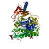
| ||||||||
|---|---|---|---|---|---|---|---|---|---|
| 1 | 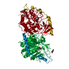
| ||||||||
| Unit cell |
|
-Components
| #1: Protein | Mass: 52038.730 Da / Num. of mol.: 1 / Source method: isolated from a natural source / Source: (natural) Homo sapiens (human) / Cellular location: LYSOSOME / Organ: PLACENTA / References: UniProt: P15289, cerebroside-sulfatase |
|---|---|
| #2: Polysaccharide | 2-acetamido-2-deoxy-beta-D-glucopyranose-(1-4)-2-acetamido-2-deoxy-beta-D-glucopyranose Source method: isolated from a genetically manipulated source |
| #3: Sugar | ChemComp-NAG /   Type: D-saccharide, beta linking / Mass: 221.208 Da / Num. of mol.: 1 Type: D-saccharide, beta linking / Mass: 221.208 Da / Num. of mol.: 1Source method: isolated from a genetically manipulated source Formula: C8H15NO6 |
| #4: Chemical | ChemComp-CA /   Mass: 40.078 Da / Num. of mol.: 1 / Source method: obtained synthetically / Formula: Ca Mass: 40.078 Da / Num. of mol.: 1 / Source method: obtained synthetically / Formula: Ca |
| #5: Water | ChemComp-HOH /  Mass: 18.015 Da / Num. of mol.: 34 / Source method: isolated from a natural source / Formula: H2O Mass: 18.015 Da / Num. of mol.: 34 / Source method: isolated from a natural source / Formula: H2O |
-Experimental details
-Experiment
| Experiment | Method: X-RAY DIFFRACTION / Number of used crystals: 1 |
|---|
- Sample preparation
| Crystal | Density Matthews: 4 Å3/Da / Density % sol: 68.99 % | ||||||||||||||||||||||||||||||||||||
|---|---|---|---|---|---|---|---|---|---|---|---|---|---|---|---|---|---|---|---|---|---|---|---|---|---|---|---|---|---|---|---|---|---|---|---|---|---|
| Crystal grow | Temperature: 293 K / Method: vapor diffusion, hanging drop / pH: 6.5 Details: cacodylate, sodium fluoride, PEG8000, pH 6.5, VAPOR DIFFUSION, HANGING DROP, temperature 293K | ||||||||||||||||||||||||||||||||||||
| Crystal grow | *PLUS Temperature: 20 ℃ / pH: 7.3 / Method: vapor diffusion, hanging drop | ||||||||||||||||||||||||||||||||||||
| Components of the solutions | *PLUS
|
-Data collection
| Diffraction | Mean temperature: 294 K |
|---|---|
| Diffraction source | Source: ROTATING ANODE / Type: RIGAKU RU200 / Wavelength: 1.5418 Å |
| Detector | Type: RIGAKU RAXIS IV / Detector: IMAGE PLATE / Date: Sep 28, 1998 / Details: mirrors |
| Radiation | Protocol: SINGLE WAVELENGTH / Monochromatic (M) / Laue (L): M / Scattering type: x-ray |
| Radiation wavelength | Wavelength: 1.5418 Å / Relative weight: 1 |
| Reflection | Num. obs: 13454 / Biso Wilson estimate: 37.3 Å2 |
| Reflection | *PLUS Highest resolution: 3.2 Å / % possible obs: 85.9 % |
- Processing
| Software |
| ||||||||||||||||||||||||||||||||||||
|---|---|---|---|---|---|---|---|---|---|---|---|---|---|---|---|---|---|---|---|---|---|---|---|---|---|---|---|---|---|---|---|---|---|---|---|---|---|
| Refinement | Method to determine structure: MOLECULAR REPLACEMENT Starting model: PDB ENTRY 1AUK Resolution: 3.2→8 Å / Rfactor Rfree error: 0.009 / Data cutoff high absF: 10000000 / Data cutoff low absF: 0.001 / Isotropic thermal model: RESTRAINED / Cross valid method: THROUGHOUT / σ(F): 2 / Stereochemistry target values: Engh & Huber
| ||||||||||||||||||||||||||||||||||||
| Displacement parameters | Biso mean: 32.3 Å2
| ||||||||||||||||||||||||||||||||||||
| Refine analyze |
| ||||||||||||||||||||||||||||||||||||
| Refinement step | Cycle: LAST / Resolution: 3.2→8 Å
| ||||||||||||||||||||||||||||||||||||
| Refine LS restraints |
| ||||||||||||||||||||||||||||||||||||
| LS refinement shell | Resolution: 3.2→3.34 Å / Rfactor Rfree error: 0.034 / Total num. of bins used: 8
| ||||||||||||||||||||||||||||||||||||
| Xplor file |
| ||||||||||||||||||||||||||||||||||||
| Refinement | *PLUS Highest resolution: 3.2 Å / Lowest resolution: 20 Å / % reflection Rfree: 6.1 % / Rfactor Rfree: 0.242 / Rfactor Rwork: 0.202 | ||||||||||||||||||||||||||||||||||||
| Solvent computation | *PLUS | ||||||||||||||||||||||||||||||||||||
| Displacement parameters | *PLUS | ||||||||||||||||||||||||||||||||||||
| Refine LS restraints | *PLUS
| ||||||||||||||||||||||||||||||||||||
| LS refinement shell | *PLUS Highest resolution: 3.2 Å / Rfactor Rfree: 0.258 / Rfactor Rwork: 0.246 |
