 Movie
Movie Controller


+ Open data

- Basic information
| Entry | Database: PDB / ID: 1auk | |||||||||
|---|---|---|---|---|---|---|---|---|---|---|
| Title | HUMAN ARYLSULFATASE A | |||||||||
 Components Components | ARYLSULFATASE A | |||||||||
 Keywords Keywords | HYDROLASE / CEREBROSIDE-3-SULFATE HYDROLYSIS / LYSOSOMAL ENZYME | |||||||||
| Function / homology |  Function and homology information Function and homology informationcerebroside-sulfatase / cerebroside-sulfatase activity / The activation of arylsulfatases / sulfuric ester hydrolase activity / arylsulfatase activity / Glycosphingolipid catabolism / lysosomal lumen / lipid metabolic process / azurophil granule lumen / lysosome ...cerebroside-sulfatase / cerebroside-sulfatase activity / The activation of arylsulfatases / sulfuric ester hydrolase activity / arylsulfatase activity / Glycosphingolipid catabolism / lysosomal lumen / lipid metabolic process / azurophil granule lumen / lysosome / endoplasmic reticulum lumen / calcium ion binding / Neutrophil degranulation / extracellular exosome / extracellular region Similarity search - Function | |||||||||
| Biological species |  Homo sapiens (human) Homo sapiens (human) | |||||||||
| Method |  X-RAY DIFFRACTION / SYNCHROTRON / MIRAS / Resolution: 2.1 Å X-RAY DIFFRACTION / SYNCHROTRON / MIRAS / Resolution: 2.1 Å | |||||||||
 Authors Authors | Lukatela, G. / Krauss, N. / Theis, K. / Gieselmann, V. / Von Figura, K. / Saenger, W. | |||||||||
 Citation Citation | Journal: Biochemistry / Year: 1998 Title: Crystal structure of human arylsulfatase A: the aldehyde function and the metal ion at the active site suggest a novel mechanism for sulfate ester hydrolysis. Authors: Lukatela, G. / Krauss, N. / Theis, K. / Selmer, T. / Gieselmann, V. / von Figura, K. / Saenger, W. | |||||||||
| History |
|
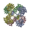
- Structure visualization
| Structure viewer | Molecule: MolmilJmol/JSmol |
|---|
- Downloads & links
-Download
| PDBx/mmCIF format | 1auk.cif.gz | 110.9 KB | Display | PDBx/mmCIF format |
|---|---|---|---|---|
| PDB format | pdb1auk.ent.gz | 82.8 KB | Display | PDB format |
| PDBx/mmJSON format | 1auk.json.gz | Tree view | PDBx/mmJSON format | |
| Others |  Other downloads Other downloads |
-Validation report
| Arichive directory | https://data.pdbj.org/pub/pdb/validation_reports/au/1aukftp://data.pdbj.org/pub/pdb/validation_reports/au/1auk | HTTPS FTP |
|---|
-Related structure data
| Similar structure data |
|---|

-Links
 PDBj
PDBj
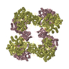
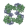
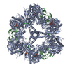
- Assembly
| Deposited unit | 
| ||||||||
|---|---|---|---|---|---|---|---|---|---|
| 1 | x 8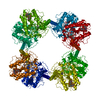
| ||||||||
| Unit cell |
|
-Components
| #1: Protein | Mass: 51956.734 Da / Num. of mol.: 1 Source method: isolated from a genetically manipulated source Source: (gene. exp.) Homo sapiens (human) / Description: C-DNA EXPRESSION / Cellular location: LYSOSOME / Gene: ARSA / Organ: TESTIS / Plasmid: PBEHCell line (production host): BABY HAMSTER KIDNEY CELLS (BHK) Cellular location (production host): SECRETED ENZYME / Production host:  Mesocricetus auratus (golden hamster) / References: UniProt: P15289, cerebroside-sulfatase Mesocricetus auratus (golden hamster) / References: UniProt: P15289, cerebroside-sulfatase |
|---|---|
| #2: Polysaccharide | 2-acetamido-2-deoxy-beta-D-glucopyranose-(1-4)-2-acetamido-2-deoxy-beta-D-glucopyranose Source method: isolated from a genetically manipulated source |
| #3: Chemical | ChemComp-MG /   Mass: 24.305 Da / Num. of mol.: 1 / Source method: obtained synthetically / Formula: Mg Mass: 24.305 Da / Num. of mol.: 1 / Source method: obtained synthetically / Formula: Mg |
| #4: Water | ChemComp-HOH /  Mass: 18.015 Da / Num. of mol.: 176 / Source method: isolated from a natural source / Formula: H2O Mass: 18.015 Da / Num. of mol.: 176 / Source method: isolated from a natural source / Formula: H2O |
| Has protein modification | Y |
-Experimental details
-Experiment
| Experiment | Method: X-RAY DIFFRACTION / Number of used crystals: 1 |
|---|
- Sample preparation
| Crystal | Density Matthews: 3.3 Å3/Da / Density % sol: 63 % | ||||||||||||||||||||||||||||||||||||||||||||||||
|---|---|---|---|---|---|---|---|---|---|---|---|---|---|---|---|---|---|---|---|---|---|---|---|---|---|---|---|---|---|---|---|---|---|---|---|---|---|---|---|---|---|---|---|---|---|---|---|---|---|
| Crystal grow | Temperature: 291 K / Method: vapor diffusion, hanging drop / pH: 5.4 Details: PROTEIN WAS CRYSTALLIZED BY VAPOR DIFFUSION IN HANGING DROPS AT 291 K. SOLUTION CONTAINING 10MG/ML PROTEIN, 10 MM TRIS/HCL (PH 7.4) AND 150 MM NACL WAS MIXED WITH SAME VOLUME OF RESERVOIR ...Details: PROTEIN WAS CRYSTALLIZED BY VAPOR DIFFUSION IN HANGING DROPS AT 291 K. SOLUTION CONTAINING 10MG/ML PROTEIN, 10 MM TRIS/HCL (PH 7.4) AND 150 MM NACL WAS MIXED WITH SAME VOLUME OF RESERVOIR SOLUTION, CONTAINING 100 MM NA-ACETATE (PH 5.0 - 5.4) AND 10 - 13 % PEG 6000, vapor diffusion - hanging drop PH range: 5.0-5.4 | ||||||||||||||||||||||||||||||||||||||||||||||||
| Crystal grow | *PLUS Temperature: 18 ℃ / Method: vapor diffusion, hanging drop | ||||||||||||||||||||||||||||||||||||||||||||||||
| Components of the solutions | *PLUS
|
-Data collection
| Diffraction | Mean temperature: 277 K |
|---|---|
| Diffraction source | Source: SYNCHROTRON / Site: SRS  / Beamline: PX9.5 / Wavelength: 0.91 / Beamline: PX9.5 / Wavelength: 0.91 |
| Detector | Type: MARRESEARCH / Detector: IMAGE PLATE / Date: Mar 1, 1996 / Details: MIRRORS |
| Radiation | Monochromatic (M) / Laue (L): M / Scattering type: x-ray |
| Radiation wavelength | Wavelength: 0.91 Å / Relative weight: 1 |
| Reflection | Resolution: 2.1→30 Å / Num. obs: 49794 / % possible obs: 96.1 % / Observed criterion σ(I): -3 / Redundancy: 6.2 % / Biso Wilson estimate: 34.97 Å2 / Rsym value: 0.055 / Net I/σ(I): 9.4 |
| Reflection shell | Resolution: 2.1→2.2 Å / Redundancy: 3.5 % / Mean I/σ(I) obs: 4.3 / Rsym value: 0.35 / % possible all: 98 |
| Reflection | *PLUS Rmerge(I) obs: 0.063 |
| Reflection shell | *PLUS % possible obs: 96.7 % / Rmerge(I) obs: 0.276 |
- Processing
| Software |
| |||||||||||||||||||||||||||||||||||||||||||||||||||||||||||||||
|---|---|---|---|---|---|---|---|---|---|---|---|---|---|---|---|---|---|---|---|---|---|---|---|---|---|---|---|---|---|---|---|---|---|---|---|---|---|---|---|---|---|---|---|---|---|---|---|---|---|---|---|---|---|---|---|---|---|---|---|---|---|---|---|---|
| Refinement | Method to determine structure: MIRAS / Resolution: 2.1→30 Å / σ(F): 0.5
| |||||||||||||||||||||||||||||||||||||||||||||||||||||||||||||||
| Displacement parameters | Biso mean: 43.1 Å2 | |||||||||||||||||||||||||||||||||||||||||||||||||||||||||||||||
| Refinement step | Cycle: LAST / Resolution: 2.1→30 Å
| |||||||||||||||||||||||||||||||||||||||||||||||||||||||||||||||
| Refine LS restraints |
| |||||||||||||||||||||||||||||||||||||||||||||||||||||||||||||||
| Software | *PLUS Name: REFMAC / Classification: refinement | |||||||||||||||||||||||||||||||||||||||||||||||||||||||||||||||
| Refinement | *PLUS Num. reflection all: 49794 | |||||||||||||||||||||||||||||||||||||||||||||||||||||||||||||||
| Solvent computation | *PLUS | |||||||||||||||||||||||||||||||||||||||||||||||||||||||||||||||
| Displacement parameters | *PLUS | |||||||||||||||||||||||||||||||||||||||||||||||||||||||||||||||
| Refine LS restraints | *PLUS
|
