+ Open data
- Basic information
| Entry | Database: PDB / ID: 1e2s | ||||||||||||
|---|---|---|---|---|---|---|---|---|---|---|---|---|---|
| Title | Crystal structure of an Arylsulfatase A mutant C69A | ||||||||||||
 Components Components | Arylsulfatase A | ||||||||||||
 Keywords Keywords | HYDROLASE / CEREBROSIDE-3-SULFATE HYDROLYSIS / LYSOSOMAL ENZYME / FORMYLGLYCINE | ||||||||||||
| Function / homology |  Function and homology information Function and homology informationcerebroside-sulfatase / cerebroside-sulfatase activity / The activation of arylsulfatases / sulfuric ester hydrolase activity / arylsulfatase activity / Glycosphingolipid catabolism / lysosomal lumen / lipid metabolic process / azurophil granule lumen / lysosome ...cerebroside-sulfatase / cerebroside-sulfatase activity / The activation of arylsulfatases / sulfuric ester hydrolase activity / arylsulfatase activity / Glycosphingolipid catabolism / lysosomal lumen / lipid metabolic process / azurophil granule lumen / lysosome / endoplasmic reticulum lumen / calcium ion binding / Neutrophil degranulation / extracellular exosome / extracellular region Similarity search - Function | ||||||||||||
| Biological species |  Homo sapiens (human) Homo sapiens (human) | ||||||||||||
| Method |  X-RAY DIFFRACTION / X-RAY DIFFRACTION /  SYNCHROTRON / SYNCHROTRON /  MOLECULAR REPLACEMENT / Resolution: 2.35 Å MOLECULAR REPLACEMENT / Resolution: 2.35 Å | ||||||||||||
 Authors Authors | von Buelow, R. / Schmidt, B. / Dierks, T. / von Figura, K. / Uson, I. | ||||||||||||
 Citation Citation |  Journal: J. Mol. Biol. / Year: 2001 Journal: J. Mol. Biol. / Year: 2001Title: Crystal structure of an enzyme-substrate complex provides insight into the interaction between human arylsulfatase A and its substrates during catalysis. Authors: von Bulow, R. / Schmidt, B. / Dierks, T. / von Figura, K. / Uson, I. | ||||||||||||
| History |
|
- Structure visualization
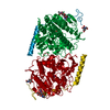
| Structure viewer | Molecule:  Molmil Molmil Jmol/JSmol Jmol/JSmol |
|---|
- Downloads & links
- Download
| PDBx/mmCIF format |  1e2s.cif.gz 1e2s.cif.gz | 112.8 KB | Display |  PDBx/mmCIF format PDBx/mmCIF format |
|---|---|---|---|---|
| PDB format |  pdb1e2s.ent.gz pdb1e2s.ent.gz | 83.3 KB | Display |  PDB format PDB format |
| PDBx/mmJSON format |  1e2s.json.gz 1e2s.json.gz | Tree view |  PDBx/mmJSON format PDBx/mmJSON format | |
| Others |  Other downloads Other downloads |
-Validation report
| Arichive directory |  https://data.pdbj.org/pub/pdb/validation_reports/e2/1e2s https://data.pdbj.org/pub/pdb/validation_reports/e2/1e2s ftp://data.pdbj.org/pub/pdb/validation_reports/e2/1e2s ftp://data.pdbj.org/pub/pdb/validation_reports/e2/1e2s | HTTPS FTP |
|---|
-Related structure data
| Related structure data | 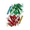 1e1zC 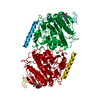 1e3cC  1aukS S: Starting model for refinement C: citing same article ( |
|---|---|
| Similar structure data |
- Links
Links
- Assembly
| Deposited unit | 
| ||||||||
|---|---|---|---|---|---|---|---|---|---|
| 1 | 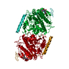
| ||||||||
| Unit cell |
|
- Components
| #1: Protein | Mass: 51926.750 Da / Num. of mol.: 1 / Mutation: C69A Source method: isolated from a genetically manipulated source Source: (gene. exp.)  Homo sapiens (human) / Cellular location: LYSOSOME / Gene: ARSA / Organ: TESTIS / Cell line (production host): MEF / Production host: Homo sapiens (human) / Cellular location: LYSOSOME / Gene: ARSA / Organ: TESTIS / Cell line (production host): MEF / Production host:  | ||
|---|---|---|---|
| #2: Polysaccharide | 2-acetamido-2-deoxy-beta-D-glucopyranose-(1-4)-2-acetamido-2-deoxy-beta-D-glucopyranose Source method: isolated from a genetically manipulated source | ||
| #3: Chemical | ChemComp-MG / | ||
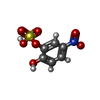
| #4: Chemical | ChemComp-CSN / | ||
| #5: Water | ChemComp-HOH / | ||
| Compound details | CHAIN P ENGINEERED| Has protein modification | Y | |
-Experimental details
-Experiment
| Experiment | Method:  X-RAY DIFFRACTION / Number of used crystals: 1 X-RAY DIFFRACTION / Number of used crystals: 1 |
|---|
- Sample preparation
| Crystal | Density Matthews: 3.3 Å3/Da / Density % sol: 63 % |
|---|---|
| Crystal grow | Temperature: 291 K / Method: vapor diffusion, hanging drop / pH: 5.4 Details: PROTEIN WAS CRYSTALLIZED BY VAPOR DIFFUSION IN HANGING DROPS AT 291 K. SOLUTION CONTAINING 10MG/ML PROTEIN, 10 MM TRIS/HCL (PH 7.4) AND 150 MM NACL WAS MIXED WITH SAME VOLUME OF RESERVOIR ...Details: PROTEIN WAS CRYSTALLIZED BY VAPOR DIFFUSION IN HANGING DROPS AT 291 K. SOLUTION CONTAINING 10MG/ML PROTEIN, 10 MM TRIS/HCL (PH 7.4) AND 150 MM NACL WAS MIXED WITH SAME VOLUME OF RESERVOIR SOLUTION, CONTAINING 100 MM NA-ACETATE (PH 5.0 - 5.4) AND 10 - 13 % PEG 6000 |
-Data collection
| Diffraction | Mean temperature: 277 K |
|---|---|
| Diffraction source | Source:  SYNCHROTRON / Site: SYNCHROTRON / Site:  EMBL/DESY, HAMBURG EMBL/DESY, HAMBURG  / Beamline: X11 / Wavelength: 0.91 / Beamline: X11 / Wavelength: 0.91 |
| Detector | Type: MARRESEARCH / Detector: IMAGE PLATE / Date: Feb 15, 1999 / Details: BENT CRYSTAL |
| Radiation | Monochromator: GE(111) / Protocol: SINGLE WAVELENGTH / Monochromatic (M) / Laue (L): M / Scattering type: x-ray |
| Radiation wavelength | Wavelength: 0.91 Å / Relative weight: 1 |
| Reflection | Resolution: 2.35→40 Å / Num. obs: 35243 / % possible obs: 99.3 % / Redundancy: 4.5 % / Biso Wilson estimate: 39.8 Å2 / Rsym value: 0.085 / Net I/σ(I): 16.9 |
| Reflection shell | Resolution: 2.35→2.45 Å / Redundancy: 4 % / Mean I/σ(I) obs: 2.8 / Rsym value: 0.34 / % possible all: 100 |
- Processing
| Software |
| ||||||||||||||||||||||||||||||||||||||||||||||||||||||||||||||||||||||||||||||||||||
|---|---|---|---|---|---|---|---|---|---|---|---|---|---|---|---|---|---|---|---|---|---|---|---|---|---|---|---|---|---|---|---|---|---|---|---|---|---|---|---|---|---|---|---|---|---|---|---|---|---|---|---|---|---|---|---|---|---|---|---|---|---|---|---|---|---|---|---|---|---|---|---|---|---|---|---|---|---|---|---|---|---|---|---|---|---|
| Refinement | Method to determine structure:  MOLECULAR REPLACEMENT MOLECULAR REPLACEMENTStarting model: 1AUK Resolution: 2.35→40 Å / SU B: 4.61 / SU ML: 0.119 / Cross valid method: FREE R-VALUE / σ(F): 0 / ESU R: 0.192 / ESU R Free: 0.129
| ||||||||||||||||||||||||||||||||||||||||||||||||||||||||||||||||||||||||||||||||||||
| Displacement parameters | Biso mean: 43.3 Å2 | ||||||||||||||||||||||||||||||||||||||||||||||||||||||||||||||||||||||||||||||||||||
| Refinement step | Cycle: LAST / Resolution: 2.35→40 Å
| ||||||||||||||||||||||||||||||||||||||||||||||||||||||||||||||||||||||||||||||||||||
| Refine LS restraints |
|
 Movie
Movie Controller





 PDBj
PDBj


