 Movie
Movie Controller

[English] 日本語
 Yorodumi
- PDB-4ytg: Crystal structure of Porphyromonas gingivalis peptidylarginine de... -
+ Open data

- Basic information
| Entry | Database: PDB / ID: 4ytg | ||||||
|---|---|---|---|---|---|---|---|
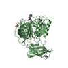
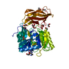
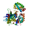
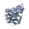
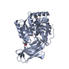
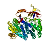
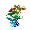
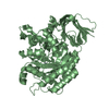
| Title | Crystal structure of Porphyromonas gingivalis peptidylarginine deiminase (PPAD) mutant C351A in complex with dipeptide Met-Arg. | ||||||
 Components Components | Peptidylarginine deiminase | ||||||
 Keywords Keywords | HYDROLASE / Peptidylarginine deiminase / citrullination | ||||||
| Function / homology |  Function and homology information Function and homology informationHydrolases; Acting on carbon-nitrogen bonds, other than peptide bonds; In linear amidines / agmatine deiminase activity / putrescine biosynthetic process / protein-arginine deiminase activity / extracellular region Similarity search - Function | ||||||
| Biological species |  Porphyromonas gingivalis (bacteria) Porphyromonas gingivalis (bacteria) | ||||||
| Method |  X-RAY DIFFRACTION / SYNCHROTRON / MOLECULAR REPLACEMENT / Resolution: 1.8 Å X-RAY DIFFRACTION / SYNCHROTRON / MOLECULAR REPLACEMENT / Resolution: 1.8 Å | ||||||
 Authors Authors | Goulas, T. / Mizgalska, D. / Garcia-Ferrer, I. / Kantyka, T. / Guevara, T. / Szmigielski, B. / Sroka, A. / Millan, C. / Uson, I. / Veillard, F. ...Goulas, T. / Mizgalska, D. / Garcia-Ferrer, I. / Kantyka, T. / Guevara, T. / Szmigielski, B. / Sroka, A. / Millan, C. / Uson, I. / Veillard, F. / Potempa, B. / Mydel, P. / Sola, M. / Potempa, J. / Gomis-Ruth, F.X. | ||||||
 Citation Citation | Journal: Sci Rep / Year: 2015 Title: Structure and mechanism of a bacterial host-protein citrullinating virulence factor, Porphyromonas gingivalis peptidylarginine deiminase. Authors: Goulas, T. / Mizgalska, D. / Garcia-Ferrer, I. / Kantyka, T. / Guevara, T. / Szmigielski, B. / Sroka, A. / Millan, C. / Uson, I. / Veillard, F. / Potempa, B. / Mydel, P. / Sola, M. / ...Authors: Goulas, T. / Mizgalska, D. / Garcia-Ferrer, I. / Kantyka, T. / Guevara, T. / Szmigielski, B. / Sroka, A. / Millan, C. / Uson, I. / Veillard, F. / Potempa, B. / Mydel, P. / Sola, M. / Potempa, J. / Gomis-Ruth, F.X. | ||||||
| History |
|
- Structure visualization
| Structure viewer | Molecule: MolmilJmol/JSmol |
|---|
- Downloads & links
-Download
| PDBx/mmCIF format | 4ytg.cif.gz | 192.9 KB | Display | PDBx/mmCIF format |
|---|---|---|---|---|
| PDB format | pdb4ytg.ent.gz | 150.6 KB | Display | PDB format |
| PDBx/mmJSON format | 4ytg.json.gz | Tree view | PDBx/mmJSON format | |
| Others |  Other downloads Other downloads |
-Validation report
| Arichive directory | https://data.pdbj.org/pub/pdb/validation_reports/yt/4ytgftp://data.pdbj.org/pub/pdb/validation_reports/yt/4ytg | HTTPS FTP |
|---|
-Related structure data
| Related structure data | 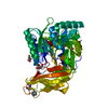 4yt9SC 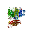 4ytbC S: Starting model for refinement C: citing same article ( |
|---|---|
| Similar structure data |

-Links
 PDBj
PDBj


- Assembly
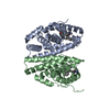
| Deposited unit | 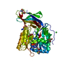
| ||||||||
|---|---|---|---|---|---|---|---|---|---|
| 1 |
| ||||||||
| Unit cell |
|
-Components
-Protein , 1 types, 1 molecules A
| #1: Protein | Mass: 48204.707 Da / Num. of mol.: 1 / Fragment: residues 44-475 / Mutation: C351A Source method: isolated from a genetically manipulated source Source: (gene. exp.) Porphyromonas gingivalis (strain ATCC BAA-308 / W83) (bacteria)Gene: PG_1424 / Production host: Porphyromonas gingivalis W83 (bacteria)References: UniProt: Q9RQJ2, Hydrolases; Acting on carbon-nitrogen bonds, other than peptide bonds; In linear amidines |
|---|
-Non-polymers , 8 types, 438 molecules 


| #2: Chemical | ChemComp-NA /  Mass: 22.990 Da / Num. of mol.: 1 / Source method: obtained synthetically / Formula: Na Mass: 22.990 Da / Num. of mol.: 1 / Source method: obtained synthetically / Formula: Na | ||||||
|---|---|---|---|---|---|---|---|
| #3: Chemical | ChemComp-CYS /  Type: L-peptide linking / Mass: 121.158 Da / Num. of mol.: 1 / Source method: obtained synthetically / Formula: C3H7NO2S Type: L-peptide linking / Mass: 121.158 Da / Num. of mol.: 1 / Source method: obtained synthetically / Formula: C3H7NO2S | ||||||
| #4: Chemical | ChemComp-MET /  Type: L-peptide linking / Mass: 149.211 Da / Num. of mol.: 1 / Source method: obtained synthetically / Formula: C5H11NO2S Type: L-peptide linking / Mass: 149.211 Da / Num. of mol.: 1 / Source method: obtained synthetically / Formula: C5H11NO2S | ||||||
| #5: Chemical | ChemComp-ARG /  Type: L-peptide linking / Mass: 175.209 Da / Num. of mol.: 1 / Source method: obtained synthetically / Formula: C6H15N4O2 Type: L-peptide linking / Mass: 175.209 Da / Num. of mol.: 1 / Source method: obtained synthetically / Formula: C6H15N4O2 | ||||||
| #6: Chemical | ChemComp-GOL /  Mass: 92.094 Da / Num. of mol.: 5 / Source method: obtained synthetically / Formula: C3H8O3 Mass: 92.094 Da / Num. of mol.: 5 / Source method: obtained synthetically / Formula: C3H8O3#7: Chemical | ChemComp-CL / |  Mass: 35.453 Da / Num. of mol.: 1 / Source method: obtained synthetically / Formula: Cl Mass: 35.453 Da / Num. of mol.: 1 / Source method: obtained synthetically / Formula: Cl#8: Chemical |  Mass: 42.020 Da / Num. of mol.: 2 / Source method: obtained synthetically / Formula: N3 Mass: 42.020 Da / Num. of mol.: 2 / Source method: obtained synthetically / Formula: N3#9: Water | ChemComp-HOH / | Mass: 18.015 Da / Num. of mol.: 426 / Source method: isolated from a natural source / Formula: H2O |
-Details
| Has protein modification | Y |
|---|
-Experimental details
-Experiment
| Experiment | Method: X-RAY DIFFRACTION |
|---|
- Sample preparation
| Crystal | Density Matthews: 2.18 Å3/Da / Density % sol: 43.47 % |
|---|---|
| Crystal grow | Temperature: 293.15 K / Method: vapor diffusion, sitting drop / pH: 6.5 Details: 100mM tri-sodium citrate, 20% [w/v] polyethylene glycol 3,000, pH5.5-6.5 PH range: 5.5-6.4 |
-Data collection
| Diffraction | Mean temperature: 100 K |
|---|---|
| Diffraction source | Source: SYNCHROTRON / Site: ALBA  / Beamline: XALOC / Wavelength: 0.9795 Å / Beamline: XALOC / Wavelength: 0.9795 Å |
| Detector | Type: DECTRIS PILATUS 6M / Detector: PIXEL / Date: Sep 21, 2014 |
| Radiation | Protocol: SINGLE WAVELENGTH / Monochromatic (M) / Laue (L): M / Scattering type: x-ray |
| Radiation wavelength | Wavelength: 0.9795 Å / Relative weight: 1 |
| Reflection | Resolution: 1.8→67.9 Å / Num. obs: 38882 / % possible obs: 99.3 % / Redundancy: 6.5 % / Biso Wilson estimate: 24.4 Å2 / Rmerge(I) obs: 0.062 / Net I/σ(I): 22.5 |
| Reflection shell | Resolution: 1.8→1.9 Å / Redundancy: 5.6 % / Rmerge(I) obs: 0.529 / Mean I/σ(I) obs: 3.7 / Num. unique all: 5771 / CC1/2: 0.927 / % possible all: 98.9 |
- Processing
| Software |
| ||||||||||||||||||||||||||||||||||||||||||||||||||||||||||||||||||||||||||||||||||||||||||||||||||||||||||||||||||
|---|---|---|---|---|---|---|---|---|---|---|---|---|---|---|---|---|---|---|---|---|---|---|---|---|---|---|---|---|---|---|---|---|---|---|---|---|---|---|---|---|---|---|---|---|---|---|---|---|---|---|---|---|---|---|---|---|---|---|---|---|---|---|---|---|---|---|---|---|---|---|---|---|---|---|---|---|---|---|---|---|---|---|---|---|---|---|---|---|---|---|---|---|---|---|---|---|---|---|---|---|---|---|---|---|---|---|---|---|---|---|---|---|---|---|---|
| Refinement | Method to determine structure: MOLECULAR REPLACEMENT Starting model: 4yt9 Resolution: 1.8→48.57 Å / Cor.coef. Fo:Fc: 0.9539 / Cor.coef. Fo:Fc free: 0.9473 / SU R Cruickshank DPI: 0.112 / Cross valid method: THROUGHOUT / σ(F): 0 / SU R Blow DPI: 0.118 / SU Rfree Blow DPI: 0.105 / SU Rfree Cruickshank DPI: 0.102
| ||||||||||||||||||||||||||||||||||||||||||||||||||||||||||||||||||||||||||||||||||||||||||||||||||||||||||||||||||
| Displacement parameters | Biso mean: 28.64 Å2
| ||||||||||||||||||||||||||||||||||||||||||||||||||||||||||||||||||||||||||||||||||||||||||||||||||||||||||||||||||
| Refine analyze | Luzzati coordinate error obs: 0.192 Å | ||||||||||||||||||||||||||||||||||||||||||||||||||||||||||||||||||||||||||||||||||||||||||||||||||||||||||||||||||
| Refinement step | Cycle: 1 / Resolution: 1.8→48.57 Å
| ||||||||||||||||||||||||||||||||||||||||||||||||||||||||||||||||||||||||||||||||||||||||||||||||||||||||||||||||||
| Refine LS restraints |
| ||||||||||||||||||||||||||||||||||||||||||||||||||||||||||||||||||||||||||||||||||||||||||||||||||||||||||||||||||
| LS refinement shell | Resolution: 1.8→1.85 Å / Total num. of bins used: 19
| ||||||||||||||||||||||||||||||||||||||||||||||||||||||||||||||||||||||||||||||||||||||||||||||||||||||||||||||||||
| Refinement TLS params. | Method: refined / Refine-ID: X-RAY DIFFRACTION
| ||||||||||||||||||||||||||||||||||||||||||||||||||||||||||||||||||||||||||||||||||||||||||||||||||||||||||||||||||
| Refinement TLS group |
|
