Hydrolases; Acting on carbon-nitrogen bonds, other than peptide bonds; In linear amidines / agmatine deiminase activity / putrescine biosynthetic process / protein-arginine deiminase activity / extracellular region Similarity search - Function
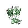
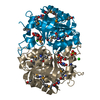
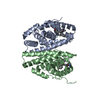
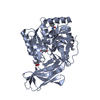
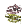
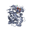
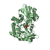
Mass: 48564.223 Da / Num. of mol.: 1 / Fragment: UNP residues 44-475 / Source method: isolated from a natural source / Source: (natural) Porphyromonas gingivalis W83 (bacteria) References: UniProt: Q9RQJ2, Hydrolases; Acting on carbon-nitrogen bonds, other than peptide bonds; In linear amidines
Protocol: SINGLE WAVELENGTH / Monochromatic (M) / Laue (L): M / Scattering type: x-ray
Radiation wavelength
Wavelength: 0.9786 Å / Relative weight: 1
Reflection
Resolution: 1.5→42.01 Å / Num. obs: 64233 / % possible obs: 98.5 % / Redundancy: 4.5 % / Biso Wilson estimate: 19.68 Å2 / Rmerge(I) obs: 0.041 / Net I/σ(I): 21
Reflection shell
Resolution: 1.5→1.59 Å / Redundancy: 4.1 % / Rmerge(I) obs: 0.19 / Mean I/σ(I) obs: 7.4 / Num. unique all: 9862 / % possible all: 95.7
-
Processing
Software
Name
Version
Classification
BUSTER
2.11.5
refinement
XDS
datareduction
XSCALE
datascaling
Arcimboldo
phasing
Refinement
Method to determine structure: AB INITIO PHASING / Resolution: 1.5→42.01 Å / Cor.coef. Fo:Fc: 0.9662 / Cor.coef. Fo:Fc free: 0.9608 / SU R Cruickshank DPI: 0.067 / Cross valid method: THROUGHOUT / σ(F): 0 / SU R Blow DPI: 0.071 / SU Rfree Blow DPI: 0.068 / SU Rfree Cruickshank DPI: 0.066
Rfactor
Num. reflection
% reflection
Selection details
Rfree
0.1771
772
1.2 %
RANDOM
Rwork
0.1569
-
-
-
obs
0.1571
64233
98.52 %
-
Displacement parameters
Biso mean: 24.44 Å2
Baniso -1
Baniso -2
Baniso -3
1-
-2.294 Å2
0 Å2
0 Å2
2-
-
2.7945 Å2
0 Å2
3-
-
-
-0.5005 Å2
Refine analyze
Luzzati coordinate error obs: 0.158 Å
Refinement step
Cycle: 1 / Resolution: 1.5→42.01 Å
Protein
Nucleic acid
Ligand
Solvent
Total
Num. atoms
3358
0
65
460
3883
Refine LS restraints
Refine-ID
Type
Dev ideal
Number
Restraint function
Weight
X-RAY DIFFRACTION
t_bond_d
0.01
3513
HARMONIC
2
X-RAY DIFFRACTION
t_angle_deg
1.03
4766
HARMONIC
2
X-RAY DIFFRACTION
t_dihedral_angle_d
1580
SINUSOIDAL
2
X-RAY DIFFRACTION
t_incorr_chiral_ct
X-RAY DIFFRACTION
t_pseud_angle
X-RAY DIFFRACTION
t_trig_c_planes
94
HARMONIC
2
X-RAY DIFFRACTION
t_gen_planes
510
HARMONIC
5
X-RAY DIFFRACTION
t_it
3513
HARMONIC
20
X-RAY DIFFRACTION
t_nbd
0
SEMIHARMONIC
5
X-RAY DIFFRACTION
t_omega_torsion
4.5
X-RAY DIFFRACTION
t_other_torsion
2.67
X-RAY DIFFRACTION
t_improper_torsion
X-RAY DIFFRACTION
t_chiral_improper_torsion
451
SEMIHARMONIC
5
X-RAY DIFFRACTION
t_sum_occupancies
6
HARMONIC
1
X-RAY DIFFRACTION
t_utility_distance
X-RAY DIFFRACTION
t_utility_angle
X-RAY DIFFRACTION
t_utility_torsion
X-RAY DIFFRACTION
t_ideal_dist_contact
4572
SEMIHARMONIC
4
LS refinement shell
Resolution: 1.5→1.54 Å / Total num. of bins used: 20
Rfactor
Num. reflection
% reflection
Rfree
0.2469
55
1.27 %
Rwork
0.1783
4289
-
all
0.1792
4344
-
obs
-
-
98.52 %
Refinement TLS params.
Method: refined / Refine-ID: X-RAY DIFFRACTION
ID
L11 (°2)
L12 (°2)
L13 (°2)
L22 (°2)
L23 (°2)
L33 (°2)
S11 (Å °)
S12 (Å °)
S13 (Å °)
S21 (Å °)
S22 (Å °)
S23 (Å °)
S31 (Å °)
S32 (Å °)
S33 (Å °)
T11 (Å2)
T12 (Å2)
T13 (Å2)
T22 (Å2)
T23 (Å2)
T33 (Å2)
Origin x (Å)
Origin y (Å)
Origin z (Å)
1
0.4687
-0.0117
0.0114
0.9799
0.0242
0.3974
-0.0095
-0.0728
0.0058
0.0679
-0.001
0.1272
-0.0237
-0.0839
0.0105
-0.0347
0.0072
0.0064
-0.0376
-0.0021
-0.0313
19.8658
35.1583
37.4495
2
0.8076
-0.1951
0.589
2.7458
-0.2787
0.3003
-0.0313
-0.1957
-0.155
0.3172
0.116
0.2974
0.0472
-0.159
-0.0848
0.0123
-0.0173
0.0839
-0.001
0.0652
-0.035
14.0604
18.586
51.9995
3
1.2007
-0.1703
-0.9392
0.7904
0.5056
2.9768
0.0727
-0.1377
-0.1431
0.0201
0.0266
-0.1682
-0.1276
0.3717
-0.0994
-0.0565
-0.0277
-0.0226
-0.0155
-0.0001
-0.0227
47.7072
30.8412
38.8953
Refinement TLS group
ID
Refine-ID
Refine TLS-ID
Selection details
1
X-RAY DIFFRACTION
1
{A|44 - 70 A|125 - 359 A|996 - 999}
2
X-RAY DIFFRACTION
2
{A|71 - 124}
3
X-RAY DIFFRACTION
3
{A|360 - 464 A|995 - 995}
+
About Yorodumi
-
News
-
Feb 9, 2022. New format data for meta-information of EMDB entries
New format data for meta-information of EMDB entries
Version 3 of the EMDB header file is now the official format.
The previous official version 1.9 will be removed from the archive.
In the structure databanks used in Yorodumi, some data are registered as the other names, "COVID-19 virus" and "2019-nCoV". Here are the details of the virus and the list of structure data.
Jan 31, 2019. EMDB accession codes are about to change! (news from PDBe EMDB page)
EMDB accession codes are about to change! (news from PDBe EMDB page)
The allocation of 4 digits for EMDB accession codes will soon come to an end. Whilst these codes will remain in use, new EMDB accession codes will include an additional digit and will expand incrementally as the available range of codes is exhausted. The current 4-digit format prefixed with “EMD-” (i.e. EMD-XXXX) will advance to a 5-digit format (i.e. EMD-XXXXX), and so on. It is currently estimated that the 4-digit codes will be depleted around Spring 2019, at which point the 5-digit format will come into force.
The EM Navigator/Yorodumi systems omit the EMD- prefix.
Related info.:Q: What is EMD? / ID/Accession-code notation in Yorodumi/EM Navigator
Yorodumi is a browser for structure data from EMDB, PDB, SASBDB, etc.
This page is also the successor to EM Navigator detail page, and also detail information page/front-end page for Omokage search.
The word "yorodu" (or yorozu) is an old Japanese word meaning "ten thousand". "mi" (miru) is to see.
Related info.:EMDB / PDB / SASBDB / Comparison of 3 databanks / Yorodumi Search / Aug 31, 2016. New EM Navigator & Yorodumi / Yorodumi Papers / Jmol/JSmol / Function and homology information / Changes in new EM Navigator and Yorodumi
 Movie
Movie Controller

 Yorodumi
 Open data
 Basic information
 Components
 Keywords
 Function and homology information
 Porphyromonas gingivalis W83 (bacteria)
 X-RAY DIFFRACTION /
  Authors
 Citation
 Structure visualization
 Downloads & links
 Other downloads
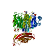


 PDBj
PDBj Assembly

 Mass: 22.990 Da / Num. of mol.: 1 / Source method: obtained synthetically / Formula: Na
 Mass: 92.094 Da / Num. of mol.: 7 / Source method: obtained synthetically / Formula: C3H8O3
 Mass: 18.015 Da / Num. of mol.: 460 / Source method: isolated from a natural source / Formula: H2O
 Sample preparation
 / Beamline: XALOC / Wavelength: 0.9786 Å
 Processing