 Movie
Movie Controller


+ Open data

- Basic information
| Entry | Database: PDB / ID: 6inh | |||||||||
|---|---|---|---|---|---|---|---|---|---|---|
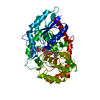
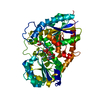
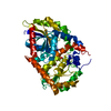
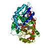
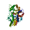
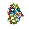
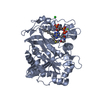
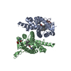
| Title | A glycosyltransferase with UDP and the substrate | |||||||||
 Components Components | UDP-glycosyltransferase 76G1 | |||||||||
 Keywords Keywords | TRANSFERASE / complex | |||||||||
| Function / homology |  Function and homology information Function and homology informationsteviolbioside glucosyltransferase activity (rebaudioside B forming) / stevioside glucosyltransferase activity (rebaudioside A forming) / quercetin 3-O-glucosyltransferase activity / quercetin 7-O-glucosyltransferase activity / UDP-glucosyltransferase activity / Transferases; Glycosyltransferases; Hexosyltransferases Similarity search - Function | |||||||||
| Biological species |  Stevia rebaudiana (plant) Stevia rebaudiana (plant) | |||||||||
| Method |  X-RAY DIFFRACTION / SYNCHROTRON / MOLECULAR REPLACEMENT / Resolution: 2.1 Å X-RAY DIFFRACTION / SYNCHROTRON / MOLECULAR REPLACEMENT / Resolution: 2.1 Å | |||||||||
 Authors Authors | Zhu, X. | |||||||||
| Funding support |  China, 2items China, 2items
| |||||||||
 Citation Citation | Journal: Nat Commun / Year: 2019 Title: Hydrophobic recognition allows the glycosyltransferase UGT76G1 to catalyze its substrate in two orientations. Authors: Yang, T. / Zhang, J. / Ke, D. / Yang, W. / Tang, M. / Jiang, J. / Cheng, G. / Li, J. / Cheng, W. / Wei, Y. / Li, Q. / Naismith, J.H. / Zhu, X. | |||||||||
| History |
|
- Structure visualization
| Structure viewer | Molecule: MolmilJmol/JSmol |
|---|
- Downloads & links
-Download
| PDBx/mmCIF format | 6inh.cif.gz | 205.7 KB | Display | PDBx/mmCIF format |
|---|---|---|---|---|
| PDB format | pdb6inh.ent.gz | 162.3 KB | Display | PDB format |
| PDBx/mmJSON format | 6inh.json.gz | Tree view | PDBx/mmJSON format | |
| Others |  Other downloads Other downloads |
-Validation report
| Arichive directory | https://data.pdbj.org/pub/pdb/validation_reports/in/6inhftp://data.pdbj.org/pub/pdb/validation_reports/in/6inh | HTTPS FTP |
|---|
-Related structure data
| Related structure data |  6infC 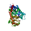 6ingC  6iniC 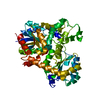 2pq6S C: citing same article ( S: Starting model for refinement |
|---|---|
| Similar structure data |


-Links
 PDBj
PDBj


- Assembly
| Deposited unit | 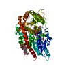
| ||||||||
|---|---|---|---|---|---|---|---|---|---|
| 1 |
| ||||||||
| Unit cell |
| ||||||||
| Components on special symmetry positions |
|
-Components
| #1: Protein | Mass: 53158.227 Da / Num. of mol.: 1 Source method: isolated from a genetically manipulated source Source: (gene. exp.) Stevia rebaudiana (plant) / Gene: UGT76G1 / Production host:  References: UniProt: Q6VAB4, Transferases; Glycosyltransferases; Hexosyltransferases | ||||
|---|---|---|---|---|---|
| #2: Chemical | ChemComp-UDP /   Type: RNA linking / Mass: 404.161 Da / Num. of mol.: 1 / Source method: obtained synthetically / Formula: C9H14N2O12P2 / Comment: UDP*YM Type: RNA linking / Mass: 404.161 Da / Num. of mol.: 1 / Source method: obtained synthetically / Formula: C9H14N2O12P2 / Comment: UDP*YM | ||||
| #3: Chemical |   Mass: 642.732 Da / Num. of mol.: 2 / Source method: obtained synthetically / Formula: C32H50O13 Mass: 642.732 Da / Num. of mol.: 2 / Source method: obtained synthetically / Formula: C32H50O13#4: Chemical |   Mass: 92.094 Da / Num. of mol.: 2 / Source method: obtained synthetically / Formula: C3H8O3 Mass: 92.094 Da / Num. of mol.: 2 / Source method: obtained synthetically / Formula: C3H8O3#5: Water | ChemComp-HOH / |  Mass: 18.015 Da / Num. of mol.: 199 / Source method: isolated from a natural source / Formula: H2O Mass: 18.015 Da / Num. of mol.: 199 / Source method: isolated from a natural source / Formula: H2O |
-Experimental details
-Experiment
| Experiment | Method: X-RAY DIFFRACTION / Number of used crystals: 1 |
|---|
- Sample preparation
| Crystal | Density Matthews: 2.35 Å3/Da / Density % sol: 47.56 % |
|---|---|
| Crystal grow | Temperature: 293 K / Method: vapor diffusion, sitting drop Details: 0.1M sodium citrate buffer at pH 5.4 and 20% PEG 4000 |
-Data collection
| Diffraction | Mean temperature: 100 K / Serial crystal experiment: N |
|---|---|
| Diffraction source | Source: SYNCHROTRON / Site: SSRF / Beamline: BL19U1 / Wavelength: 0.978 Å |
| Detector | Type: DECTRIS PILATUS 6M / Detector: PIXEL / Date: Jun 20, 2017 |
| Radiation | Protocol: SINGLE WAVELENGTH / Monochromatic (M) / Laue (L): M / Scattering type: x-ray |
| Radiation wavelength | Wavelength: 0.978 Å / Relative weight: 1 |
| Reflection | Resolution: 2.1→84.46 Å / Num. obs: 29548 / % possible obs: 100 % / Redundancy: 5.3 % / CC1/2: 0.973 / Rmerge(I) obs: 0.127 / Rpim(I) all: 0.091 / Rrim(I) all: 0.158 / Net I/σ(I): 7.2 |
| Reflection shell | Resolution: 2.1→2.15 Å / Rmerge(I) obs: 0.29 / Num. unique obs: 2159 / CC1/2: 0.916 / Rpim(I) all: 0.212 / Rrim(I) all: 0.361 |
- Processing
| Software |
| ||||||||||||||||||||||||||||||||||||||||||||||||||||||||||||||||||||||||||||||||||||||||||||||||||||||||||||||||||||||||||||||||||||||||||||||||||||||||||||||||||||||||||||||||||||||
|---|---|---|---|---|---|---|---|---|---|---|---|---|---|---|---|---|---|---|---|---|---|---|---|---|---|---|---|---|---|---|---|---|---|---|---|---|---|---|---|---|---|---|---|---|---|---|---|---|---|---|---|---|---|---|---|---|---|---|---|---|---|---|---|---|---|---|---|---|---|---|---|---|---|---|---|---|---|---|---|---|---|---|---|---|---|---|---|---|---|---|---|---|---|---|---|---|---|---|---|---|---|---|---|---|---|---|---|---|---|---|---|---|---|---|---|---|---|---|---|---|---|---|---|---|---|---|---|---|---|---|---|---|---|---|---|---|---|---|---|---|---|---|---|---|---|---|---|---|---|---|---|---|---|---|---|---|---|---|---|---|---|---|---|---|---|---|---|---|---|---|---|---|---|---|---|---|---|---|---|---|---|---|---|
| Refinement | Method to determine structure: MOLECULAR REPLACEMENT Starting model: 2PQ6 Resolution: 2.1→61.84 Å / Cor.coef. Fo:Fc: 0.971 / Cor.coef. Fo:Fc free: 0.957 / SU B: 6.853 / SU ML: 0.098 / Cross valid method: THROUGHOUT / ESU R: 0.117 / ESU R Free: 0.11 / Details: HYDROGENS HAVE BEEN ADDED IN THE RIDING POSITIONS
| ||||||||||||||||||||||||||||||||||||||||||||||||||||||||||||||||||||||||||||||||||||||||||||||||||||||||||||||||||||||||||||||||||||||||||||||||||||||||||||||||||||||||||||||||||||||
| Solvent computation | Ion probe radii: 0.9 Å / Shrinkage radii: 0.9 Å / VDW probe radii: 1.2 Å | ||||||||||||||||||||||||||||||||||||||||||||||||||||||||||||||||||||||||||||||||||||||||||||||||||||||||||||||||||||||||||||||||||||||||||||||||||||||||||||||||||||||||||||||||||||||
| Displacement parameters | Biso mean: 38.764 Å2
| ||||||||||||||||||||||||||||||||||||||||||||||||||||||||||||||||||||||||||||||||||||||||||||||||||||||||||||||||||||||||||||||||||||||||||||||||||||||||||||||||||||||||||||||||||||||
| Refinement step | Cycle: 1 / Resolution: 2.1→61.84 Å
| ||||||||||||||||||||||||||||||||||||||||||||||||||||||||||||||||||||||||||||||||||||||||||||||||||||||||||||||||||||||||||||||||||||||||||||||||||||||||||||||||||||||||||||||||||||||
| Refine LS restraints |
|