 Movie
Movie Controller

[English] 日本語
 Yorodumi
- PDB-5a68: Crystal structure of the AtTTM3 product complex with two orthopho... -
+ Open data

- Basic information
| Entry | Database: PDB / ID: 5a68 | ||||||
|---|---|---|---|---|---|---|---|
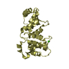
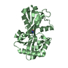
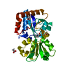
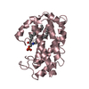
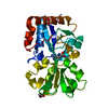
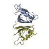
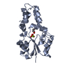
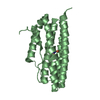
| Title | Crystal structure of the AtTTM3 product complex with two orthophosphates and manganese ions (form B) | ||||||
 Components Components | TRIPHOSPHATE TUNEL METALLOENZYME 3 | ||||||
 Keywords Keywords | HYDROLASE / INORGANIC POLYPHOSPHATE / TRIPOLYPHOSPHATE / TRIPHOSPHATE TUNNEL METALLOENZYME | ||||||
| Function / homology |  Function and homology information Function and homology informationtriphosphatase / inorganic triphosphate phosphatase activity / root development / anaphase-promoting complex / Hydrolases; Acting on acid anhydrides; In phosphorus-containing anhydrides / ATP hydrolysis activity / ATP binding / nucleus / cytoplasm Similarity search - Function | ||||||
| Biological species |  | ||||||
| Method |  X-RAY DIFFRACTION / SYNCHROTRON / OTHER / Resolution: 1.67 Å X-RAY DIFFRACTION / SYNCHROTRON / OTHER / Resolution: 1.67 Å | ||||||
 Authors Authors | Martinez, J. / Truffault, V. / Hothorn, M. | ||||||
 Citation Citation | Journal: J.Biol.Chem. / Year: 2015 Title: Structural Determinants for Substrate Binding and Catalysis in Triphosphate Tunnel Metalloenzymes. Authors: Martinez, J. / Truffault, V. / Hothorn, M. | ||||||
| History |
| ||||||
| Remark 700 | SHEET DETERMINATION METHOD: DSSP THE SHEETS PRESENTED AS "AA" IN EACH CHAIN ON SHEET RECORDS BELOW ... SHEET DETERMINATION METHOD: DSSP THE SHEETS PRESENTED AS "AA" IN EACH CHAIN ON SHEET RECORDS BELOW IS ACTUALLY AN 8-STRANDED BARREL THIS IS REPRESENTED BY A 9-STRANDED SHEET IN WHICH THE FIRST AND LAST STRANDS ARE IDENTICAL. |
- Structure visualization
| Structure viewer | Molecule: MolmilJmol/JSmol |
|---|
- Downloads & links
-Download
| PDBx/mmCIF format | 5a68.cif.gz | 104.5 KB | Display | PDBx/mmCIF format |
|---|---|---|---|---|
| PDB format | pdb5a68.ent.gz | 80.5 KB | Display | PDB format |
| PDBx/mmJSON format | 5a68.json.gz | Tree view | PDBx/mmJSON format | |
| Others |  Other downloads Other downloads |
-Validation report
| Arichive directory | https://data.pdbj.org/pub/pdb/validation_reports/a6/5a68ftp://data.pdbj.org/pub/pdb/validation_reports/a6/5a68 | HTTPS FTP |
|---|
-Related structure data
| Related structure data |  5a5yC  5a60C  5a61C  5a64C  5a65C  5a66C  5a67C C: citing same article ( |
|---|---|
| Similar structure data |
-Links
 PDBj
PDBj
- Assembly
| Deposited unit | 
| ||||||||
|---|---|---|---|---|---|---|---|---|---|
| 1 |
| ||||||||
| Unit cell |
|
-Components
| #1: Protein | Mass: 24323.764 Da / Num. of mol.: 1 Source method: isolated from a genetically manipulated source Source: (gene. exp.)  | ||||
|---|---|---|---|---|---|
| #2: Chemical |   Mass: 54.938 Da / Num. of mol.: 3 / Source method: obtained synthetically / Formula: Mn Mass: 54.938 Da / Num. of mol.: 3 / Source method: obtained synthetically / Formula: Mn#3: Chemical |   Mass: 94.971 Da / Num. of mol.: 2 / Source method: obtained synthetically / Formula: PO4 Mass: 94.971 Da / Num. of mol.: 2 / Source method: obtained synthetically / Formula: PO4#4: Water | ChemComp-HOH / |  Mass: 18.015 Da / Num. of mol.: 136 / Source method: isolated from a natural source / Formula: H2O Mass: 18.015 Da / Num. of mol.: 136 / Source method: isolated from a natural source / Formula: H2O |
-Experimental details
-Experiment
| Experiment | Method: X-RAY DIFFRACTION / Number of used crystals: 1 |
|---|
- Sample preparation
| Crystal | Density Matthews: 2.16 Å3/Da / Density % sol: 42.96 % / Description: NONE |
|---|---|
| Crystal grow | pH: 7 / Details: 0.1 M BIS TRIS PH 7.0, 20 % PEG3350 |
-Data collection
| Diffraction | Mean temperature: 100 K |
|---|---|
| Diffraction source | Source: SYNCHROTRON / Site: SLS  / Beamline: X10SA / Wavelength: 0.99998 / Beamline: X10SA / Wavelength: 0.99998 |
| Detector | Type: DECTRIS PILATUS 6M / Detector: PIXEL / Date: Feb 17, 2013 |
| Radiation | Protocol: SINGLE WAVELENGTH / Monochromatic (M) / Laue (L): M / Scattering type: x-ray |
| Radiation wavelength | Wavelength: 0.99998 Å / Relative weight: 1 |
| Reflection | Resolution: 1.67→43.2 Å / Num. obs: 23035 / % possible obs: 94.4 % / Observed criterion σ(I): -3 / Redundancy: 4.1 % / Rmerge(I) obs: 0.08 / Net I/σ(I): 9.5 |
| Reflection shell | Resolution: 1.67→1.77 Å / Redundancy: 2.95 % / Rmerge(I) obs: 0.62 / Mean I/σ(I) obs: 1.75 / % possible all: 76.8 |
- Processing
| Software |
| ||||||||||||||||||||||||||||||||||||||||||||||||||||||||||||||||||||||||||||||||||||||||||||||||||||||||||||||||||||||||||||||||||||||||||||||||||||||||||||||||||||||||||||||||||||||
|---|---|---|---|---|---|---|---|---|---|---|---|---|---|---|---|---|---|---|---|---|---|---|---|---|---|---|---|---|---|---|---|---|---|---|---|---|---|---|---|---|---|---|---|---|---|---|---|---|---|---|---|---|---|---|---|---|---|---|---|---|---|---|---|---|---|---|---|---|---|---|---|---|---|---|---|---|---|---|---|---|---|---|---|---|---|---|---|---|---|---|---|---|---|---|---|---|---|---|---|---|---|---|---|---|---|---|---|---|---|---|---|---|---|---|---|---|---|---|---|---|---|---|---|---|---|---|---|---|---|---|---|---|---|---|---|---|---|---|---|---|---|---|---|---|---|---|---|---|---|---|---|---|---|---|---|---|---|---|---|---|---|---|---|---|---|---|---|---|---|---|---|---|---|---|---|---|---|---|---|---|---|---|---|
| Refinement | Method to determine structure: OTHER Starting model: NONE Resolution: 1.67→43.2 Å / Cor.coef. Fo:Fc: 0.966 / Cor.coef. Fo:Fc free: 0.943 / SU B: 6.345 / SU ML: 0.09 / Cross valid method: THROUGHOUT / ESU R: 0.177 / ESU R Free: 0.111 / Stereochemistry target values: MAXIMUM LIKELIHOOD Details: HYDROGENS HAVE BEEN ADDED IN THE RIDING POSITIONS. U VALUES WITH TLS ADDED
| ||||||||||||||||||||||||||||||||||||||||||||||||||||||||||||||||||||||||||||||||||||||||||||||||||||||||||||||||||||||||||||||||||||||||||||||||||||||||||||||||||||||||||||||||||||||
| Solvent computation | Ion probe radii: 0.8 Å / Shrinkage radii: 0.8 Å / VDW probe radii: 1.2 Å / Solvent model: MASK | ||||||||||||||||||||||||||||||||||||||||||||||||||||||||||||||||||||||||||||||||||||||||||||||||||||||||||||||||||||||||||||||||||||||||||||||||||||||||||||||||||||||||||||||||||||||
| Displacement parameters | Biso mean: 29.197 Å2
| ||||||||||||||||||||||||||||||||||||||||||||||||||||||||||||||||||||||||||||||||||||||||||||||||||||||||||||||||||||||||||||||||||||||||||||||||||||||||||||||||||||||||||||||||||||||
| Refinement step | Cycle: LAST / Resolution: 1.67→43.2 Å
| ||||||||||||||||||||||||||||||||||||||||||||||||||||||||||||||||||||||||||||||||||||||||||||||||||||||||||||||||||||||||||||||||||||||||||||||||||||||||||||||||||||||||||||||||||||||
| Refine LS restraints |
|