 Movie
Movie Controller

[English] 日本語
 Yorodumi
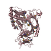
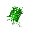
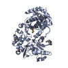
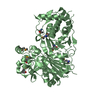
- PDB-5a5f: CRYSTAL STRUCTURE OF MURD LIGASE FROM ESCHERICHIA COLI IN COMPLEX... -
+ Open data

- Basic information
| Entry | Database: PDB / ID: 5a5f | ||||||
|---|---|---|---|---|---|---|---|
| Title | CRYSTAL STRUCTURE OF MURD LIGASE FROM ESCHERICHIA COLI IN COMPLEX WITH UMA AND ADP | ||||||
 Components Components | UDP-N-ACETYLMURAMOYLALANINE--D-GLUTAMATE LIGASE | ||||||
 Keywords Keywords | LIGASE / PEPTIDOGLYCAN SYNTHESIS / ADP-FORMING ENZYME / CELL WALL / CELL SHAPE / CELL CYCLE / NUCLEOTIDE-BINDING / ATP- BINDING / CELL DIVISION / LIGAND / CONFORMATION | ||||||
| Function / homology |  Function and homology information Function and homology informationUDP-N-acetylmuramoyl-L-alanine-D-glutamate ligase / UDP-N-acetylmuramoylalanine-D-glutamate ligase activity / peptidoglycan biosynthetic process / cell wall organization / regulation of cell shape / cell division / ATP binding / identical protein binding / cytoplasm Similarity search - Function | ||||||
| Biological species |  | ||||||
| Method |  X-RAY DIFFRACTION / SYNCHROTRON / MOLECULAR REPLACEMENT / Resolution: 1.9 Å X-RAY DIFFRACTION / SYNCHROTRON / MOLECULAR REPLACEMENT / Resolution: 1.9 Å | ||||||
 Authors Authors | Sink, R. / Kotnik, M. / Zega, A. / Barreteau, H. / Gobec, S. / Blanot, D. / Dessen, A. / Contreras-Martel, C. | ||||||
 Citation Citation | Journal: PLoS ONE / Year: 2016 Title: Crystallographic Study of Peptidoglycan Biosynthesis Enzyme MurD: Domain Movement Revisited. Authors: Sink, R. / Kotnik, M. / Zega, A. / Barreteau, H. / Gobec, S. / Blanot, D. / Dessen, A. / Contreras-Martel, C. | ||||||
| History |
|
- Structure visualization
| Structure viewer | Molecule: MolmilJmol/JSmol |
|---|
- Downloads & links
-Download
| PDBx/mmCIF format | 5a5f.cif.gz | 184.3 KB | Display | PDBx/mmCIF format |
|---|---|---|---|---|
| PDB format | pdb5a5f.ent.gz | 146.5 KB | Display | PDB format |
| PDBx/mmJSON format | 5a5f.json.gz | Tree view | PDBx/mmJSON format | |
| Others |  Other downloads Other downloads |
-Validation report
| Arichive directory | https://data.pdbj.org/pub/pdb/validation_reports/a5/5a5fftp://data.pdbj.org/pub/pdb/validation_reports/a5/5a5f | HTTPS FTP |
|---|
-Related structure data
| Related structure data | 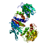 5a5eC 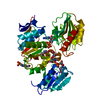 3uagS C: citing same article ( S: Starting model for refinement |
|---|---|
| Similar structure data |


-Links
 PDBj
PDBj- Assembly
| Deposited unit | 
| ||||||||
|---|---|---|---|---|---|---|---|---|---|
| 1 |
| ||||||||
| Unit cell |
|
-Components
| #1: Protein | Mass: 46976.332 Da / Num. of mol.: 1 Source method: isolated from a genetically manipulated source Source: (gene. exp.) References: UniProt: P14900, UDP-N-acetylmuramoyl-L-alanine-D-glutamate ligase | ||||
|---|---|---|---|---|---|
| #2: Chemical | ChemComp-UMA / 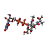  Type: L-peptide linking / Mass: 750.494 Da / Num. of mol.: 1 / Source method: obtained synthetically / Formula: C23H36N4O20P2 Type: L-peptide linking / Mass: 750.494 Da / Num. of mol.: 1 / Source method: obtained synthetically / Formula: C23H36N4O20P2 | ||||
| #3: Chemical | ChemComp-ADP /   Mass: 427.201 Da / Num. of mol.: 1 / Source method: obtained synthetically / Formula: C10H15N5O10P2 / Comment: ADP, energy-carrying molecule*YM Mass: 427.201 Da / Num. of mol.: 1 / Source method: obtained synthetically / Formula: C10H15N5O10P2 / Comment: ADP, energy-carrying molecule*YM | ||||
| #4: Chemical |   Mass: 102.046 Da / Num. of mol.: 2 / Source method: obtained synthetically / Formula: C3H2O4 Mass: 102.046 Da / Num. of mol.: 2 / Source method: obtained synthetically / Formula: C3H2O4#5: Water | ChemComp-HOH / |  Mass: 18.015 Da / Num. of mol.: 269 / Source method: isolated from a natural source / Formula: H2O Mass: 18.015 Da / Num. of mol.: 269 / Source method: isolated from a natural source / Formula: H2OHas protein modification | Y | |
-Experimental details
-Experiment
| Experiment | Method: X-RAY DIFFRACTION / Number of used crystals: 1 |
|---|
- Sample preparation
| Crystal | Density Matthews: 3.37 Å3/Da / Density % sol: 63.55 % / Description: NONE |
|---|---|
| Crystal grow | pH: 7.4 Details: 4 MG/ML PROTEIN, 1 MM UMA, 5 MM AMP-PNP, 1 MM NAN3, 1 MM DTT, 20 MM HEPES, PH 7.4, 1.8 M NA-MALONATE |
-Data collection
| Diffraction | Mean temperature: 100 K |
|---|---|
| Diffraction source | Source: SYNCHROTRON / Site: ESRF  / Beamline: ID29 / Wavelength: 0.97623 / Beamline: ID29 / Wavelength: 0.97623 |
| Detector | Type: ADSC CCD / Detector: CCD / Date: Apr 30, 2008 |
| Radiation | Protocol: SINGLE WAVELENGTH / Monochromatic (M) / Laue (L): M / Scattering type: x-ray |
| Radiation wavelength | Wavelength: 0.97623 Å / Relative weight: 1 |
| Reflection | Resolution: 1.9→46.46 Å / Num. obs: 51890 / % possible obs: 98.2 % / Observed criterion σ(I): 3 / Redundancy: 5.4 % / Biso Wilson estimate: 37.009 Å2 / Rmerge(I) obs: 0.06 / Net I/σ(I): 23.75 |
| Reflection shell | Resolution: 1.9→2.01 Å / Redundancy: 5.5 % / Rmerge(I) obs: 0.62 / Mean I/σ(I) obs: 3.22 / % possible all: 94.8 |
- Processing
| Software |
| ||||||||||||||||||||||||||||||||||||||||||||||||||||||||||||||||||||||||||||||||||||||||||||||||||||||||||||||||||||||||||||||||||||||||||||||||||||||||||||||||||||||||||||||||||||||
|---|---|---|---|---|---|---|---|---|---|---|---|---|---|---|---|---|---|---|---|---|---|---|---|---|---|---|---|---|---|---|---|---|---|---|---|---|---|---|---|---|---|---|---|---|---|---|---|---|---|---|---|---|---|---|---|---|---|---|---|---|---|---|---|---|---|---|---|---|---|---|---|---|---|---|---|---|---|---|---|---|---|---|---|---|---|---|---|---|---|---|---|---|---|---|---|---|---|---|---|---|---|---|---|---|---|---|---|---|---|---|---|---|---|---|---|---|---|---|---|---|---|---|---|---|---|---|---|---|---|---|---|---|---|---|---|---|---|---|---|---|---|---|---|---|---|---|---|---|---|---|---|---|---|---|---|---|---|---|---|---|---|---|---|---|---|---|---|---|---|---|---|---|---|---|---|---|---|---|---|---|---|---|---|
| Refinement | Method to determine structure: MOLECULAR REPLACEMENT Starting model: PDB ENTRY 3UAG Resolution: 1.9→46.47 Å / Cor.coef. Fo:Fc: 0.96 / Cor.coef. Fo:Fc free: 0.944 / SU B: 6.549 / SU ML: 0.1 / Cross valid method: THROUGHOUT / ESU R: 0.126 / ESU R Free: 0.124 / Stereochemistry target values: MAXIMUM LIKELIHOOD / Details: HYDROGENS HAVE BEEN ADDED IN THE RIDING POSITIONS.
| ||||||||||||||||||||||||||||||||||||||||||||||||||||||||||||||||||||||||||||||||||||||||||||||||||||||||||||||||||||||||||||||||||||||||||||||||||||||||||||||||||||||||||||||||||||||
| Solvent computation | Ion probe radii: 0.8 Å / Shrinkage radii: 0.8 Å / VDW probe radii: 1.2 Å / Solvent model: BABINET MODEL WITH MASK | ||||||||||||||||||||||||||||||||||||||||||||||||||||||||||||||||||||||||||||||||||||||||||||||||||||||||||||||||||||||||||||||||||||||||||||||||||||||||||||||||||||||||||||||||||||||
| Displacement parameters | Biso mean: 35.207 Å2
| ||||||||||||||||||||||||||||||||||||||||||||||||||||||||||||||||||||||||||||||||||||||||||||||||||||||||||||||||||||||||||||||||||||||||||||||||||||||||||||||||||||||||||||||||||||||
| Refinement step | Cycle: LAST / Resolution: 1.9→46.47 Å
| ||||||||||||||||||||||||||||||||||||||||||||||||||||||||||||||||||||||||||||||||||||||||||||||||||||||||||||||||||||||||||||||||||||||||||||||||||||||||||||||||||||||||||||||||||||||
| Refine LS restraints |
|