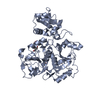
 Movie
Movie Controller


+ Open data

- Basic information
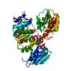
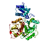
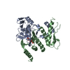
| Entry | Database: PDB / ID: 3uag | ||||||
|---|---|---|---|---|---|---|---|
| Title | UDP-N-ACETYLMURAMOYL-L-ALANINE:D-GLUTAMATE LIGASE | ||||||
 Components Components | PROTEIN (UDP-N-ACETYLMURAMOYL-L-ALANINE:D-GLUTAMATE LIGASE) | ||||||
 Keywords Keywords | LIGASE / PEPTIDOGLYCAN SYNTHESIS / MURD / ADP-FORMING ENZYME | ||||||
| Function / homology |  Function and homology information Function and homology informationUDP-N-acetylmuramoyl-L-alanine-D-glutamate ligase / UDP-N-acetylmuramoylalanine-D-glutamate ligase activity / peptidoglycan biosynthetic process / cell wall organization / regulation of cell shape / cell division / ATP binding / identical protein binding / cytoplasm Similarity search - Function | ||||||
| Biological species |  | ||||||
| Method |  X-RAY DIFFRACTION / SYNCHROTRON / OTHER / Resolution: 1.77 Å X-RAY DIFFRACTION / SYNCHROTRON / OTHER / Resolution: 1.77 Å | ||||||
 Authors Authors | Bertrand, J.A. / Auger, G. / Martin, L. / Fanchon, E. / Blanot, D. / Le Beller, D. / Van Heijenoort, J. / Dideberg, O. | ||||||
 Citation Citation | Journal: J.Mol.Biol. / Year: 1999 Title: Determination of the MurD mechanism through crystallographic analysis of enzyme complexes. Authors: Bertrand, J.A. / Auger, G. / Martin, L. / Fanchon, E. / Blanot, D. / Le Beller, D. / van Heijenoort, J. / Dideberg, O. #1: Journal: Protein Expr.Purif. / Year: 1998 Title: Large-scale preparation, purification, and crystallization of UDP-N-acetylmuramoyl-L-alanine: D-glutamate ligase from Escherichia coli. Authors: Auger, G. / Martin, L. / Bertrand, J. / Ferrari, P. / Fanchon, E. / Vaganay, S. / Petillot, Y. / van Heijenoort, J. / Blanot, D. / Dideberg, O. #2: Journal: Embo J. / Year: 1997Title: Crystal structure of UDP-N-acetylmuramoyl-L-alanine:D-glutamate ligase from Escherichia coli. Authors: Bertrand, J.A. / Auger, G. / Fanchon, E. / Martin, L. / Blanot, D. / van Heijenoort, J. / Dideberg, O. | ||||||
| History |
|
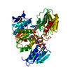
- Structure visualization
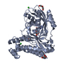
| Structure viewer | Molecule: MolmilJmol/JSmol |
|---|
- Downloads & links
-Download
| PDBx/mmCIF format | 3uag.cif.gz | 107.4 KB | Display | PDBx/mmCIF format |
|---|---|---|---|---|
| PDB format | pdb3uag.ent.gz | 79.6 KB | Display | PDB format |
| PDBx/mmJSON format | 3uag.json.gz | Tree view | PDBx/mmJSON format | |
| Others |  Other downloads Other downloads |
-Validation report
| Arichive directory | https://data.pdbj.org/pub/pdb/validation_reports/ua/3uagftp://data.pdbj.org/pub/pdb/validation_reports/ua/3uag | HTTPS FTP |
|---|
-Related structure data


-Links
 PDBj
PDBj- Assembly
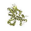
| Deposited unit | 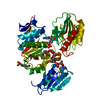
| ||||||||
|---|---|---|---|---|---|---|---|---|---|
| 1 |
| ||||||||
| Unit cell |
|
-Components
-Protein , 1 types, 1 molecules A
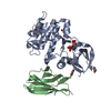
| #1: Protein | Mass: 46932.254 Da / Num. of mol.: 1 Source method: isolated from a genetically manipulated source Details: UMA, ADP & MANGANESE BOUND, PH 7.2 / Source: (gene. exp.) References: UniProt: P14900, UDP-N-acetylmuramoyl-L-alanine-D-glutamate ligase |
|---|
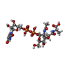
-Non-polymers , 5 types, 363 molecules 


| #2: Chemical | ChemComp-MN /  Mass: 54.938 Da / Num. of mol.: 1 / Source method: obtained synthetically / Formula: Mn Mass: 54.938 Da / Num. of mol.: 1 / Source method: obtained synthetically / Formula: Mn |
|---|---|
| #3: Chemical | ChemComp-UMA /  Type: L-peptide linking / Mass: 750.494 Da / Num. of mol.: 1 / Source method: obtained synthetically / Formula: C23H36N4O20P2 Type: L-peptide linking / Mass: 750.494 Da / Num. of mol.: 1 / Source method: obtained synthetically / Formula: C23H36N4O20P2 |
| #4: Chemical | ChemComp-ADP /  Mass: 427.201 Da / Num. of mol.: 1 / Source method: obtained synthetically / Formula: C10H15N5O10P2 / Comment: ADP, energy-carrying molecule*YM Mass: 427.201 Da / Num. of mol.: 1 / Source method: obtained synthetically / Formula: C10H15N5O10P2 / Comment: ADP, energy-carrying molecule*YM |
| #5: Chemical | ChemComp-EPE /  Mass: 238.305 Da / Num. of mol.: 1 / Source method: obtained synthetically / Formula: C8H18N2O4S / Comment: pH buffer*YM Mass: 238.305 Da / Num. of mol.: 1 / Source method: obtained synthetically / Formula: C8H18N2O4S / Comment: pH buffer*YM |
| #6: Water | ChemComp-HOH / Mass: 18.015 Da / Num. of mol.: 359 / Source method: isolated from a natural source / Formula: H2O |
-Details
| Compound details | RESIDUE 198 IS A MODIFIED LYSINE WHICH IS CARBAMYLATED AT THE ZETA-AMINO GROUP. THE CARBAMYLATED ...RESIDUE 198 IS A MODIFIED LYSINE WHICH IS CARBAMYLAT | ||
|---|---|---|---|
| Nonpolymer details | RESIDUE 198 IS A MODIFIED LYSINE WHICH IS CARBAMYLAT| Sequence details | KCX 198, MODIFIED LYSINE RESIDUE | |
-Experimental details
-Experiment
| Experiment | Method: X-RAY DIFFRACTION / Number of used crystals: 1 |
|---|
- Sample preparation
| Crystal | Density Matthews: 3.01 Å3/Da / Density % sol: 59.62 % | ||||||||||||||||||||||||||||||||||||||||||||||||
|---|---|---|---|---|---|---|---|---|---|---|---|---|---|---|---|---|---|---|---|---|---|---|---|---|---|---|---|---|---|---|---|---|---|---|---|---|---|---|---|---|---|---|---|---|---|---|---|---|---|
| Crystal grow | pH: 7.2 / Details: pH 7.2 | ||||||||||||||||||||||||||||||||||||||||||||||||
| Crystal grow | *PLUS pH: 7.5 / Method: vapor diffusionDetails: drop consists of equal volume of protein and reservoir solutions | ||||||||||||||||||||||||||||||||||||||||||||||||
| Components of the solutions | *PLUS
|
-Data collection
| Diffraction | Mean temperature: 100 K |
|---|---|
| Diffraction source | Source: SYNCHROTRON / Site: EMBL/DESY, HAMBURG  / Beamline: BW7B / Wavelength: 0.881 / Beamline: BW7B / Wavelength: 0.881 |
| Detector | Type: MARRESEARCH / Detector: IMAGE PLATE / Date: Sep 1, 1996 |
| Radiation | Protocol: SINGLE WAVELENGTH / Monochromatic (M) / Laue (L): M / Scattering type: x-ray |
| Radiation wavelength | Wavelength: 0.881 Å / Relative weight: 1 |
| Reflection | Resolution: 1.77→65.9 Å / Num. obs: 54268 / % possible obs: 99.2 % / Redundancy: 4.6 % / Rsym value: 0.066 / Net I/σ(I): 25.6 |
| Reflection shell | Resolution: 1.77→1.83 Å / Redundancy: 4.4 % / Mean I/σ(I) obs: 8.6 / Rsym value: 0.204 / % possible all: 98.7 |
| Reflection | *PLUS Rmerge(I) obs: 0.066 |
| Reflection shell | *PLUS % possible obs: 98.7 % / Rmerge(I) obs: 0.204 |
- Processing
| Software |
| ||||||||||||||||||||||||||||||||||||||||||||||||||||||||||||
|---|---|---|---|---|---|---|---|---|---|---|---|---|---|---|---|---|---|---|---|---|---|---|---|---|---|---|---|---|---|---|---|---|---|---|---|---|---|---|---|---|---|---|---|---|---|---|---|---|---|---|---|---|---|---|---|---|---|---|---|---|---|
| Refinement | Method to determine structure: OTHER / Resolution: 1.77→8 Å / Rfactor Rfree error: 0.004 / Data cutoff high absF: 0 / Data cutoff low absF: 0 / Cross valid method: THROUGHOUT / σ(F): 0
| ||||||||||||||||||||||||||||||||||||||||||||||||||||||||||||
| Displacement parameters | Biso mean: 16.91 Å2
| ||||||||||||||||||||||||||||||||||||||||||||||||||||||||||||
| Refinement step | Cycle: LAST / Resolution: 1.77→8 Å
| ||||||||||||||||||||||||||||||||||||||||||||||||||||||||||||
| Refine LS restraints |
| ||||||||||||||||||||||||||||||||||||||||||||||||||||||||||||
| LS refinement shell | Resolution: 1.77→1.85 Å / Rfactor Rfree error: 0.013 / Total num. of bins used: 8
| ||||||||||||||||||||||||||||||||||||||||||||||||||||||||||||
| Xplor file | Serial no: 1 / Param file: PARHCSDX.PRO / Topol file: TOPHCSDX.PRO | ||||||||||||||||||||||||||||||||||||||||||||||||||||||||||||
| Software | *PLUS Name: X-PLOR / Version: 3.1 / Classification: refinement | ||||||||||||||||||||||||||||||||||||||||||||||||||||||||||||
| Refine LS restraints | *PLUS
|
