 Movie
Movie Controller

[English] 日本語
 Yorodumi
- PDB-3u07: Crystal Structure of the VPA0106 protein from Vibrio parahaemolyt... -
+ Open data

- Basic information
| Entry | Database: PDB / ID: 3u07 | ||||||
|---|---|---|---|---|---|---|---|
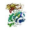
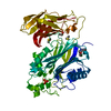
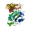
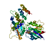
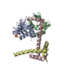
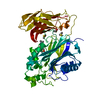
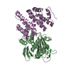
| Title | Crystal Structure of the VPA0106 protein from Vibrio parahaemolyticus. Northeast Structural Genomics Consortium Target VpR106. | ||||||
 Components Components | uncharacterized protein VPA0106 | ||||||
 Keywords Keywords | Structural Genomics / Unknown Function / PSI-Biology / Protein Structure Initiative / NESG / Northeast Structural Genomics Consortium | ||||||
| Function / homology |  Function and homology information Function and homology informationJelly Rolls - #1600 / Domain of unknown function DUF1254 / Domain of unknown function DUF1254 superfamily / Protein of unknown function (DUF1254) / Domain of unknown function DUF1214 / Protein of unknown function (DUF1214) / Jelly Rolls / Sandwich / Mainly Beta Similarity search - Domain/homology | ||||||
| Biological species |   Vibrio parahaemolyticus (bacteria) Vibrio parahaemolyticus (bacteria) | ||||||
| Method |  X-RAY DIFFRACTION / SYNCHROTRON / SAD / Resolution: 2.083 Å X-RAY DIFFRACTION / SYNCHROTRON / SAD / Resolution: 2.083 Å | ||||||
 Authors Authors | Vorobiev, S. / Neely, H. / Seetharaman, J. / Patel, P. / Xiao, R. / Ciccosanti, C. / Wang, H. / Everett, J.K. / Nair, R. / Acton, T.B. ...Vorobiev, S. / Neely, H. / Seetharaman, J. / Patel, P. / Xiao, R. / Ciccosanti, C. / Wang, H. / Everett, J.K. / Nair, R. / Acton, T.B. / Rost, B. / Montelione, G.T. / Tong, L. / Hunt, J.F. / Northeast Structural Genomics Consortium (NESG) | ||||||
 Citation Citation | Journal: To be Published Title: Crystal Structure of the VPA0106 protein from Vibrio parahaemolyticus. Authors: Vorobiev, S. / Neely, H. / Seetharaman, J. / Patel, P. / Xiao, R. / Ciccosanti, C. / Wang, H. / Everett, J.K. / Nair, R. / Acton, T.B. / Rost, B. / Montelione, G.T. / Tong, L. / Hunt, J.F. | ||||||
| History |
|
- Structure visualization
| Structure viewer | Molecule: MolmilJmol/JSmol |
|---|
- Downloads & links
-Download
| PDBx/mmCIF format | 3u07.cif.gz | 471 KB | Display | PDBx/mmCIF format |
|---|---|---|---|---|
| PDB format | pdb3u07.ent.gz | 385.6 KB | Display | PDB format |
| PDBx/mmJSON format | 3u07.json.gz | Tree view | PDBx/mmJSON format | |
| Others |  Other downloads Other downloads |
-Validation report
| Arichive directory | https://data.pdbj.org/pub/pdb/validation_reports/u0/3u07ftp://data.pdbj.org/pub/pdb/validation_reports/u0/3u07 | HTTPS FTP |
|---|
-Related structure data
| Similar structure data | |
|---|---|
| Other databases |


-Links
 PDBj
PDBj- Assembly
| Deposited unit | 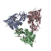
| ||||||||
|---|---|---|---|---|---|---|---|---|---|
| 1 | 
| ||||||||
| 2 | 
| ||||||||
| 3 | 
| ||||||||
| Unit cell |
| ||||||||
| Details | monomer,55.15 kD,94.5% |
-Components
| #1: Protein | Mass: 51150.629 Da / Num. of mol.: 3 / Mutation: R60E, I64R Source method: isolated from a genetically manipulated source Source: (gene. exp.) Vibrio parahaemolyticus (bacteria) / Strain: RIMD 2210633 / Serotype O3:K6 / Gene: VPA0106 / Plasmid: pET21_NESG, VpR106-RM.2 / Production host: #2: Water | ChemComp-HOH / |  Mass: 18.015 Da / Num. of mol.: 780 / Source method: isolated from a natural source / Formula: H2O Mass: 18.015 Da / Num. of mol.: 780 / Source method: isolated from a natural source / Formula: H2OHas protein modification | Y | |
|---|
-Experimental details
-Experiment
| Experiment | Method: X-RAY DIFFRACTION / Number of used crystals: 1 |
|---|
- Sample preparation
| Crystal | Density Matthews: 2.79 Å3/Da / Density % sol: 55.95 % |
|---|---|
| Crystal grow | Temperature: 291 K / Method: microbatch crystallization under oil / pH: 6 Details: 20% PEG 4000, 0.1M sodium nitrate, 0.1M MES, pH 6.0, Microbatch crystallization under oil, temperature 291K |
-Data collection
| Diffraction | Mean temperature: 100 K |
|---|---|
| Diffraction source | Source: SYNCHROTRON / Site: NSLS  / Beamline: X4A / Wavelength: 0.97898 Å / Beamline: X4A / Wavelength: 0.97898 Å |
| Detector | Type: ADSC QUANTUM 4 / Detector: CCD / Date: Sep 18, 2011 |
| Radiation | Monochromator: Si 111 CHANNEL / Protocol: SINGLE WAVELENGTH / Monochromatic (M) / Laue (L): M / Scattering type: x-ray |
| Radiation wavelength | Wavelength: 0.97898 Å / Relative weight: 1 |
| Reflection | Resolution: 2.083→50 Å / Num. all: 198295 / Num. obs: 188380 / % possible obs: 95.6 % / Observed criterion σ(F): 0 / Observed criterion σ(I): 0 / Redundancy: 1.4 % / Biso Wilson estimate: 25.05 Å2 / Rmerge(I) obs: 0.048 / Net I/σ(I): 13.3 |
| Reflection shell | Resolution: 2.1→2.18 Å / Redundancy: 1.4 % / Rmerge(I) obs: 0.205 / Mean I/σ(I) obs: 2.8 / Num. unique all: 19654 / % possible all: 79.8 |
- Processing
| Software |
| |||||||||||||||||||||||||||||||||||||||||||||||||||||||||||||||||||||||||||||||||||||||||||||||||||||||||||||||||||||||||||||||||||||||||||||||||||||||||||||||||||||||||||||||||||||||||||||||||||||||||||||||||||||||||
|---|---|---|---|---|---|---|---|---|---|---|---|---|---|---|---|---|---|---|---|---|---|---|---|---|---|---|---|---|---|---|---|---|---|---|---|---|---|---|---|---|---|---|---|---|---|---|---|---|---|---|---|---|---|---|---|---|---|---|---|---|---|---|---|---|---|---|---|---|---|---|---|---|---|---|---|---|---|---|---|---|---|---|---|---|---|---|---|---|---|---|---|---|---|---|---|---|---|---|---|---|---|---|---|---|---|---|---|---|---|---|---|---|---|---|---|---|---|---|---|---|---|---|---|---|---|---|---|---|---|---|---|---|---|---|---|---|---|---|---|---|---|---|---|---|---|---|---|---|---|---|---|---|---|---|---|---|---|---|---|---|---|---|---|---|---|---|---|---|---|---|---|---|---|---|---|---|---|---|---|---|---|---|---|---|---|---|---|---|---|---|---|---|---|---|---|---|---|---|---|---|---|---|---|---|---|---|---|---|---|---|---|---|---|---|---|---|---|---|
| Refinement | Method to determine structure: SAD / Resolution: 2.083→46.958 Å / Occupancy max: 1 / Occupancy min: 0.36 / SU ML: 0.6 / Isotropic thermal model: Isotropic / Cross valid method: THROUGHOUT / σ(F): 1.11 / Phase error: 21.96 / Stereochemistry target values: ML
| |||||||||||||||||||||||||||||||||||||||||||||||||||||||||||||||||||||||||||||||||||||||||||||||||||||||||||||||||||||||||||||||||||||||||||||||||||||||||||||||||||||||||||||||||||||||||||||||||||||||||||||||||||||||||
| Solvent computation | Shrinkage radii: 0.9 Å / VDW probe radii: 1.11 Å / Solvent model: FLAT BULK SOLVENT MODEL / Bsol: 34.226 Å2 / ksol: 0.351 e/Å3 | |||||||||||||||||||||||||||||||||||||||||||||||||||||||||||||||||||||||||||||||||||||||||||||||||||||||||||||||||||||||||||||||||||||||||||||||||||||||||||||||||||||||||||||||||||||||||||||||||||||||||||||||||||||||||
| Displacement parameters | Biso max: 124.28 Å2 / Biso mean: 32.622 Å2 / Biso min: 1.71 Å2
| |||||||||||||||||||||||||||||||||||||||||||||||||||||||||||||||||||||||||||||||||||||||||||||||||||||||||||||||||||||||||||||||||||||||||||||||||||||||||||||||||||||||||||||||||||||||||||||||||||||||||||||||||||||||||
| Refinement step | Cycle: LAST / Resolution: 2.083→46.958 Å
| |||||||||||||||||||||||||||||||||||||||||||||||||||||||||||||||||||||||||||||||||||||||||||||||||||||||||||||||||||||||||||||||||||||||||||||||||||||||||||||||||||||||||||||||||||||||||||||||||||||||||||||||||||||||||
| Refine LS restraints |
| |||||||||||||||||||||||||||||||||||||||||||||||||||||||||||||||||||||||||||||||||||||||||||||||||||||||||||||||||||||||||||||||||||||||||||||||||||||||||||||||||||||||||||||||||||||||||||||||||||||||||||||||||||||||||
| LS refinement shell | Refine-ID: X-RAY DIFFRACTION / Total num. of bins used: 30
| |||||||||||||||||||||||||||||||||||||||||||||||||||||||||||||||||||||||||||||||||||||||||||||||||||||||||||||||||||||||||||||||||||||||||||||||||||||||||||||||||||||||||||||||||||||||||||||||||||||||||||||||||||||||||
| Refinement TLS params. | Method: refined / Refine-ID: X-RAY DIFFRACTION
| |||||||||||||||||||||||||||||||||||||||||||||||||||||||||||||||||||||||||||||||||||||||||||||||||||||||||||||||||||||||||||||||||||||||||||||||||||||||||||||||||||||||||||||||||||||||||||||||||||||||||||||||||||||||||
| Refinement TLS group |
|
