- PDB-3lvr: The crystal structure of ASAP3 in complex with Arf6 in transition... -
+
Open data
ID or keywords:
Loading...
-
Basic information
Entry
Database: PDB / ID: 3lvr
Title
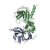
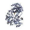
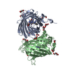
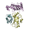
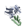
The crystal structure of ASAP3 in complex with Arf6 in transition state soaked with Calcium
Components
Arf-GAP with SH3 domain, ANK repeat and PH domain-containing protein 3, ADP-ribosylation factor 6
Keywords
PROTEIN TRANSPORT / Arf6 / ASAP3 / UPLC1 / GDP / Calcium / ArfGAP / Arf / Linkers / Alternative splicing / ANK repeat / Coiled coil / Cytoplasm / Metal-binding / Phosphoprotein / Polymorphism / Zinc / Zinc-finger / Cell membrane / Endosome / ER-Golgi transport / Golgi apparatus / GTP-binding / Lipoprotein / Myristate / Nucleotide-binding / Transport
Function / homology
Function and homology information
erythrocyte apoptotic process / maintenance of postsynaptic density structure / protein localization to cleavage furrow / positive regulation of mitotic cytokinetic process / establishment of epithelial cell polarity / regulation of dendritic spine development / negative regulation of protein localization to cell surface / protein localization to endosome / regulation of Rac protein signal transduction / negative regulation of dendrite development ...erythrocyte apoptotic process / maintenance of postsynaptic density structure / protein localization to cleavage furrow / positive regulation of mitotic cytokinetic process / establishment of epithelial cell polarity / regulation of dendritic spine development / negative regulation of protein localization to cell surface / protein localization to endosome / regulation of Rac protein signal transduction / negative regulation of dendrite development / ruffle assembly / regulation of filopodium assembly / positive regulation of keratinocyte migration / positive regulation of focal adhesion disassembly / endocytic recycling / MET receptor recycling / thioesterase binding / Flemming body / regulation of stress fiber assembly / filopodium membrane / TBC/RABGAPs / protein localization to cell surface / cortical actin cytoskeleton organization / hepatocyte apoptotic process / positive regulation of actin filament polymerization / cleavage furrow / positive regulation of GTPase activity / endocytic vesicle / regulation of presynapse assembly / synaptic vesicle endocytosis / ruffle / vesicle-mediated transport / signaling adaptor activity / GTPase activator activity / small monomeric GTPase / protein localization to plasma membrane / positive regulation of protein localization to plasma membrane / positive regulation of protein secretion / liver development / negative regulation of receptor-mediated endocytosis / cellular response to nerve growth factor stimulus / positive regulation of neuron projection development / intracellular protein transport / recycling endosome membrane / GDP binding / nervous system development / cell migration / Clathrin-mediated endocytosis / presynapse / G protein activity / early endosome membrane / midbody / cell cortex / cell differentiation / cell adhesion / endosome / postsynapse / cell division / focal adhesion / GTPase activity / GTP binding / glutamatergic synapse / Golgi apparatus / extracellular exosome / zinc ion binding / nucleoplasm / membrane / plasma membrane / cytoplasm / cytosol Similarity search - Function
Serine Threonine Protein Phosphatase 5, Tetratricopeptide repeat - #950 / Arf GTPase activating protein, ASAP3 / : / ASAP, PH domain / Arf-GAP with SH3 domain, ANK repeat and PH domain-containing protein / Arf GTPase activating protein / ADP-ribosylation factor 6 / BAR domain of APPL family / Arf GTPase activating protein / ArfGAP domain superfamily ...Serine Threonine Protein Phosphatase 5, Tetratricopeptide repeat - #950 / Arf GTPase activating protein, ASAP3 / : / ASAP, PH domain / Arf-GAP with SH3 domain, ANK repeat and PH domain-containing protein / Arf GTPase activating protein / ADP-ribosylation factor 6 / BAR domain of APPL family / Arf GTPase activating protein / ArfGAP domain superfamily / Putative GTPase activating protein for Arf / ARF GTPase-activating proteins domain profile. / Putative GTP-ase activating proteins for the small GTPase, ARF / ARFGAP/RecO-like zinc finger / BAR domain / Small GTPase superfamily, ARF type / Annexin V; domain 1 / Small GTPase Arf domain profile. / Sar1p-like members of the Ras-family of small GTPases / Small GTPase superfamily, ARF/SAR type / ADP-ribosylation factor family / AH/BAR domain superfamily / ARF-like small GTPases; ARF, ADP-ribosylation factor / Ankyrin repeat-containing domain / PH domain / PH domain profile. / Pleckstrin homology domain. / Pleckstrin homology domain / Ankyrin repeat profile. / Ankyrin repeats (3 copies) / Ankyrin repeat region circular profile. / ankyrin repeats / Ankyrin repeat / Rab subfamily of small GTPases / Ankyrin repeat-containing domain superfamily / Serine Threonine Protein Phosphatase 5, Tetratricopeptide repeat / Alpha Horseshoe / Small GTP-binding protein domain / PH-like domain superfamily / P-loop containing nucleotide triphosphate hydrolases / Rossmann fold / P-loop containing nucleoside triphosphate hydrolase / Orthogonal Bundle / 3-Layer(aba) Sandwich / Mainly Alpha / Alpha Beta Similarity search - Domain/homology
ALUMINUM FLUORIDE / GUANOSINE-5'-DIPHOSPHATE / ADP-ribosylation factor 6 / Arf-GAP with SH3 domain, ANK repeat and PH domain-containing protein 3 Similarity search - Component
Arf-GAPwithSH3domain, ANKrepeatandPHdomain-containingprotein3, ADP-ribosylationfactor6 / ASAP3 / Development and differentiation-enhancing factor-like 1 / Protein up-regulated in liver ...ASAP3 / Development and differentiation-enhancing factor-like 1 / Protein up-regulated in liver cancer 1 / Arf6 / UPLC1
Mass: 54825.930 Da / Num. of mol.: 1 Fragment: GAP and Ankyrin domain, residues 416-697, residues 11-175 Source method: isolated from a genetically manipulated source Details: Missing the N-terminus amphipathic helix for Arf6 Source: (gene. exp.) Homo sapiens (human), (gene. exp.) synthetic construct (others) Gene: ASAP3, ARF6 / Production host: Escherichia coli (E. coli) / References: UniProt: Q8TDY4, PDB-3LVQ, UniProt: P62330
Resolution: 3.38→29.46 Å / Cor.coef. Fo:Fc: 0.94 / Cor.coef. Fo:Fc free: 0.901 / SU B: 56.231 / SU ML: 0.451 / Cross valid method: THROUGHOUT / ESU R Free: 0.61 / Stereochemistry target values: MAXIMUM LIKELIHOOD / Details: HYDROGENS HAVE BEEN ADDED IN THE RIDING POSITIONS
Rfactor
Num. reflection
% reflection
Selection details
Rfree
0.28129
437
5 %
RANDOM
Rwork
0.20746
-
-
-
obs
0.21095
8294
100 %
-
Solvent computation
Ion probe radii: 0.8 Å / Shrinkage radii: 0.8 Å / VDW probe radii: 1.2 Å / Solvent model: MASK
Displacement parameters
Biso mean: 125.397 Å2
Baniso -1
Baniso -2
Baniso -3
1-
-6.75 Å2
-3.38 Å2
0 Å2
2-
-
-6.75 Å2
0 Å2
3-
-
-
10.13 Å2
Refinement step
Cycle: LAST / Resolution: 3.38→29.46 Å
Protein
Nucleic acid
Ligand
Solvent
Total
Num. atoms
3257
0
35
6
3298
Refine LS restraints
Refine-ID
Type
Dev ideal
Dev ideal target
Number
X-RAY DIFFRACTION
r_bond_refined_d
0.006
0.022
3348
X-RAY DIFFRACTION
r_angle_refined_deg
1.058
1.977
4540
X-RAY DIFFRACTION
r_dihedral_angle_1_deg
5.473
5
415
X-RAY DIFFRACTION
r_dihedral_angle_2_deg
38.465
24.211
152
X-RAY DIFFRACTION
r_dihedral_angle_3_deg
17.901
15
575
X-RAY DIFFRACTION
r_dihedral_angle_4_deg
13.86
15
25
X-RAY DIFFRACTION
r_chiral_restr
0.076
0.2
513
X-RAY DIFFRACTION
r_gen_planes_refined
0.002
0.02
2509
X-RAY DIFFRACTION
r_nbd_refined
0.201
0.2
1544
X-RAY DIFFRACTION
r_nbtor_refined
0.299
0.2
2257
X-RAY DIFFRACTION
r_xyhbond_nbd_refined
0.137
0.2
114
X-RAY DIFFRACTION
r_metal_ion_refined
0.113
0.2
2
X-RAY DIFFRACTION
r_symmetry_vdw_refined
0.195
0.2
27
X-RAY DIFFRACTION
r_mcbond_it
0.223
1.5
2129
X-RAY DIFFRACTION
r_mcangle_it
0.405
2
3329
X-RAY DIFFRACTION
r_scbond_it
0.446
3
1375
X-RAY DIFFRACTION
r_scangle_it
0.813
4.5
1211
LS refinement shell
Resolution: 3.381→3.467 Å / Total num. of bins used: 20
Rfactor
Num. reflection
% reflection
Rfree
0.296
32
-
Rwork
0.249
601
-
obs
-
601
100 %
Refinement TLS params.
Method: refined / Refine-ID: X-RAY DIFFRACTION
ID
L11 (°2)
L12 (°2)
L13 (°2)
L22 (°2)
L23 (°2)
L33 (°2)
S11 (Å °)
S12 (Å °)
S13 (Å °)
S21 (Å °)
S22 (Å °)
S23 (Å °)
S31 (Å °)
S32 (Å °)
S33 (Å °)
T11 (Å2)
T12 (Å2)
T13 (Å2)
T22 (Å2)
T23 (Å2)
T33 (Å2)
Origin x (Å)
Origin y (Å)
Origin z (Å)
1
5.3368
1.1971
-0.0031
9.7407
-0.8969
4.9919
-0.1094
0.0863
-0.7944
0.0645
0.1212
-0.4716
0.7445
-0.4123
-0.0118
-0.5114
0.104
0.1519
-0.3132
-0.1904
-0.4966
37.709
31.5561
-3.219
2
11.3975
0.2885
0.3194
7.8783
0.0237
5.7939
-0.2783
-0.3039
0.8954
0.3346
0.0096
0.8382
-0.1207
-0.2749
0.2687
-0.71
0.0865
0.1309
-0.6942
-0.1082
-0.4774
20.5949
55.7745
5.3018
Refinement TLS group
ID
Refine-ID
Refine TLS-ID
Auth asym-ID
Auth seq-ID
1
X-RAY DIFFRACTION
1
E
423 - 679
2
X-RAY DIFFRACTION
2
A
11 - 174
+
About Yorodumi
-
News
-
Feb 9, 2022. New format data for meta-information of EMDB entries
New format data for meta-information of EMDB entries
Version 3 of the EMDB header file is now the official format.
The previous official version 1.9 will be removed from the archive.
In the structure databanks used in Yorodumi, some data are registered as the other names, "COVID-19 virus" and "2019-nCoV". Here are the details of the virus and the list of structure data.
Jan 31, 2019. EMDB accession codes are about to change! (news from PDBe EMDB page)
EMDB accession codes are about to change! (news from PDBe EMDB page)
The allocation of 4 digits for EMDB accession codes will soon come to an end. Whilst these codes will remain in use, new EMDB accession codes will include an additional digit and will expand incrementally as the available range of codes is exhausted. The current 4-digit format prefixed with “EMD-” (i.e. EMD-XXXX) will advance to a 5-digit format (i.e. EMD-XXXXX), and so on. It is currently estimated that the 4-digit codes will be depleted around Spring 2019, at which point the 5-digit format will come into force.
The EM Navigator/Yorodumi systems omit the EMD- prefix.
Related info.:Q: What is EMD? / ID/Accession-code notation in Yorodumi/EM Navigator
Yorodumi is a browser for structure data from EMDB, PDB, SASBDB, etc.
This page is also the successor to EM Navigator detail page, and also detail information page/front-end page for Omokage search.
The word "yorodu" (or yorozu) is an old Japanese word meaning "ten thousand". "mi" (miru) is to see.
Related info.:EMDB / PDB / SASBDB / Comparison of 3 databanks / Yorodumi Search / Aug 31, 2016. New EM Navigator & Yorodumi / Yorodumi Papers / Jmol/JSmol / Function and homology information / Changes in new EM Navigator and Yorodumi
 Movie
Movie Controller

 Yorodumi
 Open data
 Basic information
 Components
 Keywords
 Function and homology information
 Homo sapiens (human)
 X-RAY DIFFRACTION /
  Authors
 Citation
 Structure visualization
 Downloads & links
 Other downloads
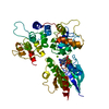


 PDBj
PDBj


 Assembly
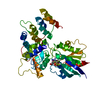


 Mass: 65.409 Da / Num. of mol.: 1 / Source method: obtained synthetically / Formula: Zn
 Mass: 24.305 Da / Num. of mol.: 1 / Source method: obtained synthetically / Formula: Mg
 Type: RNA linking / Mass: 443.201 Da / Num. of mol.: 1 / Source method: obtained synthetically / Formula: C10H15N5O11P2 / Comment: GDP, energy-carrying molecule*YM
 Mass: 83.977 Da / Num. of mol.: 1 / Source method: obtained synthetically / Formula: AlF3
 Mass: 40.078 Da / Num. of mol.: 1 / Source method: obtained synthetically / Formula: Ca
 Sample preparation
 / Beamline: X06SA / Wavelength: 1.3838 Å
 Processing