| Entry | Database: PDB / ID: 6mib
|
|---|
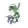
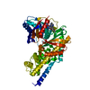
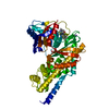
| Title | Crystal structure of the ILK ATP-binding deficient mutant (L207W)/alpha-parvin core complex |
|---|
 Components Components | - Alpha-parvin
- Integrin-linked protein kinase
|
|---|
 Keywords Keywords | CELL ADHESION / Pseudokinase / Protein complex |
|---|
| Function / homology |  Function and homology information Function and homology information
actin-mediated cell contraction / smooth muscle cell chemotaxis / establishment or maintenance of cell polarity regulating cell shape / protein localization to cell cortex / caveola assembly / Regulation of cytoskeletal remodeling and cell spreading by IPP complex components / Localization of the PINCH-ILK-PARVIN complex to focal adhesions / negative regulation of neural precursor cell proliferation / positive regulation of signal transduction / fibroblast migration ...actin-mediated cell contraction / smooth muscle cell chemotaxis / establishment or maintenance of cell polarity regulating cell shape / protein localization to cell cortex / caveola assembly / Regulation of cytoskeletal remodeling and cell spreading by IPP complex components / Localization of the PINCH-ILK-PARVIN complex to focal adhesions / negative regulation of neural precursor cell proliferation / positive regulation of signal transduction / fibroblast migration / nerve development / myelination in peripheral nervous system / establishment or maintenance of epithelial cell apical/basal polarity / Cell-extracellular matrix interactions / outflow tract septum morphogenesis / positive regulation of BMP signaling pathway / sprouting angiogenesis / cell projection organization / neural precursor cell proliferation / heterotypic cell-cell adhesion / branching involved in ureteric bud morphogenesis / outflow tract morphogenesis / protein kinase inhibitor activity / establishment or maintenance of cell polarity / cilium assembly / positive regulation of osteoblast differentiation / substrate adhesion-dependent cell spreading / positive regulation of substrate adhesion-dependent cell spreading / integrin-mediated signaling pathway / cell-matrix adhesion / mitotic spindle organization / tumor necrosis factor-mediated signaling pathway / sarcomere / phosphatidylinositol 3-kinase/protein kinase B signal transduction / platelet aggregation / integrin binding / cell morphogenesis / Z disc / positive regulation of canonical Wnt signaling pathway / lamellipodium / actin cytoskeleton / actin cytoskeleton organization / actin binding / cell cortex / protein-macromolecule adaptor activity / positive regulation of canonical NF-kappaB signal transduction / protein kinase activity / cell differentiation / protein stabilization / cadherin binding / signaling receptor binding / focal adhesion / centrosome / positive regulation of cell population proliferation / protein kinase binding / positive regulation of DNA-templated transcription / chromatin / magnesium ion binding / nucleoplasm / ATP binding / membrane / nucleus / plasma membrane / cytosol / cytoplasmSimilarity search - Function Integrin-linked protein kinase, pseudokinase domain / Parvin / Calponin-like domain / Actin-binding Protein, T-fimbrin; domain 1 / Calponin homology domain / Calponin homology (CH) domain / : / Calponin homology domain / CH domain superfamily / Calponin homology (CH) domain profile. ...Integrin-linked protein kinase, pseudokinase domain / Parvin / Calponin-like domain / Actin-binding Protein, T-fimbrin; domain 1 / Calponin homology domain / Calponin homology (CH) domain / : / Calponin homology domain / CH domain superfamily / Calponin homology (CH) domain profile. / Ankyrin repeat profile. / Ankyrin repeats (3 copies) / Ankyrin repeat region circular profile. / ankyrin repeats / Ankyrin repeat / Ankyrin repeat-containing domain superfamily / Protein tyrosine and serine/threonine kinase / Serine-threonine/tyrosine-protein kinase, catalytic domain / Phosphorylase Kinase; domain 1 / Phosphorylase Kinase; domain 1 / Transferase(Phosphotransferase) domain 1 / Transferase(Phosphotransferase); domain 1 / Protein kinase domain profile. / Protein kinase domain / Protein kinase-like domain superfamily / 2-Layer Sandwich / Orthogonal Bundle / Mainly Alpha / Alpha BetaSimilarity search - Domain/homology |
|---|
| Biological species |  Homo sapiens (human) Homo sapiens (human) |
|---|
| Method |  X-RAY DIFFRACTION / SYNCHROTRON / MOLECULAR REPLACEMENT / Resolution: 1.8 Å X-RAY DIFFRACTION / SYNCHROTRON / MOLECULAR REPLACEMENT / Resolution: 1.8 Å |
|---|
 Authors Authors | Fukuda, K. / Qin, J. |
|---|
| Funding support |  United States, 1items United States, 1items | Organization | Grant number | Country |
|---|
| National Institutes of Health/National Human Genome Research Institute (NIH/NHGRI) | | United States |
|
|---|
 Citation Citation | Journal: Nat Commun / Year: 2018
Title: Non-catalytic signaling by pseudokinase ILK for regulating cell adhesion.
Authors: Vaynberg, J. / Fukuda, K. / Lu, F. / Bialkowska, K. / Chen, Y. / Plow, E.F. / Qin, J. |
|---|
| History | | Deposition | Sep 19, 2018 | Deposition site: RCSB / Processing site: RCSB |
|---|
| Revision 1.0 | Nov 7, 2018 | Provider: repository / Type: Initial release |
|---|
| Revision 1.1 | Dec 18, 2019 | Group: Author supporting evidence / Category: pdbx_audit_support / Item: _pdbx_audit_support.funding_organization |
|---|
| Revision 1.2 | Mar 13, 2024 | Group: Data collection / Database references / Category: chem_comp_atom / chem_comp_bond / database_2
Item: _database_2.pdbx_DOI / _database_2.pdbx_database_accession |
|---|
|
|---|
 Movie
Movie Controller

 Yorodumi
 Open data
 Basic information
 Structure visualization
 Downloads & links
 Other downloads
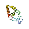


 PDBj
PDBj Assembly
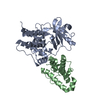

 Mass: 18.015 Da / Num. of mol.: 223 / Source method: isolated from a natural source / Formula: H2O
 Sample preparation
 Processing