Mass: 18.015 Da / Num. of mol.: 117 / Source method: isolated from a natural source / Formula: H2O
Sequence details
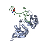
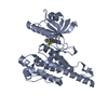
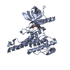
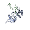
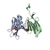
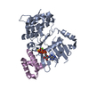
DOMAIN CONTAINING RESIDUES 19 TO 266
-
Experimental details
-
Experiment
Experiment
Method: X-RAY DIFFRACTION / Number of used crystals: 1
-
Sample preparation
Crystal
Density Matthews: 2.2 Å3/Da / Density % sol: 43 % Description: ANISOTROPIC DIFFRACTION RESULTED IN APPROXIMATELY TWENTY PERCENT OF A SINGLE WEDGE OF DATA OF BEING VERY POOR DIFFRACTION QUALITY.
Monochromator: SI (111) DOUBLE CRYSTAL MONOCHROMATOR / Protocol: SINGLE WAVELENGTH / Monochromatic (M) / Laue (L): M / Scattering type: x-ray
Radiation wavelength
Wavelength: 0.9464 Å / Relative weight: 1
Reflection
Resolution: 2→68.8 Å / Num. obs: 31249 / % possible obs: 96 % / Observed criterion σ(I): 2 / Redundancy: 2.2 % / Rmerge(I) obs: 0.12 / Net I/σ(I): 5.8
Reflection shell
Resolution: 2→2.05 Å / Redundancy: 2 % / Rmerge(I) obs: 0.58 / Mean I/σ(I) obs: 1.5 / % possible all: 82
-
Processing
Software
Name
Version
Classification
REFMAC
5.8.0049
refinement
XDS
datareduction
Aimless
datascaling
REFMAC
phasing
Refinement
Method to determine structure: MOLECULAR REPLACEMENT / Resolution: 2→68.82 Å / Cor.coef. Fo:Fc: 0.93 / Cor.coef. Fo:Fc free: 0.895 / SU B: 12.684 / SU ML: 0.172 / Cross valid method: THROUGHOUT / ESU R: 0.231 / ESU R Free: 0.199 / Stereochemistry target values: MAXIMUM LIKELIHOOD Details: HYDROGENS HAVE BEEN ADDED IN THE RIDING POSITIONS. R AND FREER FACTOR VALUES ARE LOWER THAN EXPECTED FOR TWO ANGSTROM DATA. THIS IS MOST LIKELY DUE TO A WEDGE POOR OF ANISOTROPIC DIFFRACTED ...Details: HYDROGENS HAVE BEEN ADDED IN THE RIDING POSITIONS. R AND FREER FACTOR VALUES ARE LOWER THAN EXPECTED FOR TWO ANGSTROM DATA. THIS IS MOST LIKELY DUE TO A WEDGE POOR OF ANISOTROPIC DIFFRACTED DATA BUT THE DENSITY LOOKS GOOD THROUGHOUT THE STRUCTURE.
Rfactor
Num. reflection
% reflection
Selection details
Rfree
0.26854
1579
5.1 %
RANDOM
Rwork
0.21712
-
-
-
obs
0.21979
29670
96.38 %
-
Solvent computation
Ion probe radii: 0.8 Å / Shrinkage radii: 0.8 Å / VDW probe radii: 1.2 Å / Solvent model: MASK
 Movie
Movie Controller

 Yorodumi
 Open data
 Basic information
 Components
 Keywords
 Function and homology information
 HOMO SAPIENS (human)
 X-RAY DIFFRACTION /
  Authors
 Citation
 Structure visualization
 Downloads & links
 Other downloads


 PDBj
PDBj


 Assembly
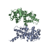

 Mass: 18.015 Da / Num. of mol.: 117 / Source method: isolated from a natural source / Formula: H2O
 Sample preparation
 / Beamline: I04 / Wavelength: 0.9464
  Processing