 Movie
Movie Controller

[English] 日本語
 Yorodumi
- PDB-6gf6: Molecular basis of egg coat filament cross-linking: high-resoluti... -
+ Open data

- Basic information
| Entry | Database: PDB / ID: 6gf6 | |||||||||||||||
|---|---|---|---|---|---|---|---|---|---|---|---|---|---|---|---|---|
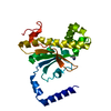
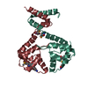
| Title | Molecular basis of egg coat filament cross-linking: high-resolution structure of the partially deglycosylated ZP1 ZP-N1 domain homodimer | |||||||||||||||
 Components Components | Zona pellucida sperm-binding protein 1,Zona pellucida sperm-binding protein 1 | |||||||||||||||
 Keywords Keywords | CELL ADHESION / Zona pellucida / ZP1 / ZP-N domain / ZP module / ZP domain / egg coat filament cross-linking / egg coat penetration by sperm | |||||||||||||||
| Function / homology |  Function and homology information Function and homology information | |||||||||||||||
| Biological species |  | |||||||||||||||
| Method |  X-RAY DIFFRACTION / SYNCHROTRON / MOLECULAR REPLACEMENT / Resolution: 2.3 Å X-RAY DIFFRACTION / SYNCHROTRON / MOLECULAR REPLACEMENT / Resolution: 2.3 Å | |||||||||||||||
 Authors Authors | Nishimura, K. / Jovine, L. | |||||||||||||||
| Funding support |  Sweden, 4items Sweden, 4items
| |||||||||||||||
 Citation Citation | Journal: Nat Commun / Year: 2019 Title: Molecular basis of egg coat cross-linking sheds light on ZP1-associated female infertility. Authors: Nishimura, K. / Dioguardi, E. / Nishio, S. / Villa, A. / Han, L. / Matsuda, T. / Jovine, L. #1: Journal: J. Mol. Biol. / Year: 1985 Title: Mouse egg extracellular coat is a matrix of interconnected filaments possessing a structural repeat. Authors: Greve, J.M. / Wassarman, P.M. #2: Journal: Development / Year: 1999 Title: Abnormal zonae pellucidae in mice lacking ZP1 result in early embryonic loss. Authors: Rankin, T. / Talbot, P. / Lee, E. / Dean, J. #3: Journal: Biol. Reprod. / Year: 2001 Title: Morphological and biochemical changes of isolated chicken egg-envelope during sperm penetration: degradation of the 97-kilodalton glycoprotein is involved in sperm-driven hole formation on the egg-envelope. Authors: Takeuchi, Y. / Cho, R. / Iwata, Y. / Nishimura, K. / Kato, T. / Aoki, N. / Kitajima, K. / Matsuda, T. #4: Journal: Biochem J / Year: 2004 Title: A newly identified zona pellucida glycoprotein, ZPD, and dimeric ZP1 of chicken egg envelope are involved in sperm activation on sperm-egg interaction. Authors: Hiroki Okumura / Yoshinori Kohno / Yuki Iwata / Hitoshi Mori / Naohito Aoki / Chihiro Sato / Ken Kitajima / Daita Nadano / Tsukasa Matsuda /  Abstract: Fertilization begins with interaction between the sperm and the egg. The surface of the vertebrate oocyte is covered with the egg envelope, which is composed of ZP (zona pellucida) glycoproteins. We ...Fertilization begins with interaction between the sperm and the egg. The surface of the vertebrate oocyte is covered with the egg envelope, which is composed of ZP (zona pellucida) glycoproteins. We have identified two glycoproteins, ZP1/gp97 and ZPC/gp42, as the major components of the chicken egg envelope. In the present study, another 42 kDa protein, designated ZPD, has been found as a new major component of the chicken egg envelope. ZPD was specifically released from the egg envelope by ultrasonication treatment without urea. ZPD cDNA was cloned using a chicken granulosa cell cDNA pool. The deduced amino acid sequence showed that preproprotein of ZPD is composed of 418 amino acid residues with four potential N-glycosylation sites and includes a ZP domain, common in vertebrate ZP glycoproteins, and a transmembrane domain. ZPD belongs phylogenetically to a distinct group from known ZP glycoprotein subfamilies, ZPA, ZPB, and ZPC. In two-dimensional gel electrophoresis ZPD proteins were identified to be several isoforms with different pI values between 5 and 7. ZP1, ZPC and the newly identified ZPD were confirmed to be the major components of chicken egg envelope by MS of proteolytic digests of whole egg envelope. The in vitro incubation of chicken sperm with calcium ionophore A23187 induced sperm activation, resulting in the fragmentation and release of a 41 kDa PNA (peanut agglutinin)-positive glycoprotein and the decrease or loss of sperm PNA-stainability. The incubation with ZPD and dimeric ZP1, but not ZPC and monomeric ZP1, also induced the decrease or loss of sperm PNA-stainability, suggesting the in vitro sperm activation by these ZP components. Collectively, ZPD might bind loosely to egg envelope matrix and play a key role in the sperm activation on avian sperm-egg interaction. #5: Journal: N. Engl. J. Med. / Year: 2014 Title: Mutant ZP1 in familial infertility. Authors: Huang, H.L. / Lv, C. / Zhao, Y.C. / Li, W. / He, X.M. / Li, P. / Sha, A.G. / Tian, X. / Papasian, C.J. / Deng, H.W. / Lu, G.X. / Xiao, H.M. #6: Journal: FEBS Open Bio / Year: 2015 Title: Identification of distinctive interdomain interactions among ZP-N, ZP-C and other domains of zona pellucida glycoproteins underlying association of chicken egg-coat matrix. Authors: Okumura, H. / Sato, T. / Sakuma, R. / Fukushima, H. / Matsuda, T. / Ujita, M. | |||||||||||||||
| History |
|
- Structure visualization
| Structure viewer | Molecule: MolmilJmol/JSmol |
|---|
- Downloads & links
-Download
| PDBx/mmCIF format | 6gf6.cif.gz | 107.5 KB | Display | PDBx/mmCIF format |
|---|---|---|---|---|
| PDB format | pdb6gf6.ent.gz | 81.2 KB | Display | PDB format |
| PDBx/mmJSON format | 6gf6.json.gz | Tree view | PDBx/mmJSON format | |
| Others |  Other downloads Other downloads |
-Validation report
| Arichive directory | https://data.pdbj.org/pub/pdb/validation_reports/gf/6gf6ftp://data.pdbj.org/pub/pdb/validation_reports/gf/6gf6 | HTTPS FTP |
|---|
-Related structure data


-Links
 PDBj
PDBj- Assembly
| Deposited unit | 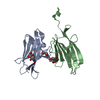
| ||||||||
|---|---|---|---|---|---|---|---|---|---|
| 1 |
| ||||||||
| Unit cell |
|
-Components
| #1: Protein | Mass: 15485.485 Da / Num. of mol.: 2 / Mutation: N121Q, G140H(6), G149S Source method: isolated from a genetically manipulated source Source: (gene. exp.)  Homo sapiens (human) / References: UniProt: A0A140JXP0 Homo sapiens (human) / References: UniProt: A0A140JXP0#2: Sugar |   Type: D-saccharide, beta linking / Mass: 221.208 Da / Num. of mol.: 2 / Source method: isolated from a natural source / Formula: C8H15NO6 Type: D-saccharide, beta linking / Mass: 221.208 Da / Num. of mol.: 2 / Source method: isolated from a natural source / Formula: C8H15NO6#3: Chemical | ChemComp-GOL / |   Mass: 92.094 Da / Num. of mol.: 1 / Source method: obtained synthetically / Formula: C3H8O3 Mass: 92.094 Da / Num. of mol.: 1 / Source method: obtained synthetically / Formula: C3H8O3#4: Water | ChemComp-HOH / |  Mass: 18.015 Da / Num. of mol.: 88 / Source method: isolated from a natural source / Formula: H2O Mass: 18.015 Da / Num. of mol.: 88 / Source method: isolated from a natural source / Formula: H2OHas protein modification | Y | |
|---|
-Experimental details
-Experiment
| Experiment | Method: X-RAY DIFFRACTION / Number of used crystals: 1 |
|---|
- Sample preparation
| Crystal | Density Matthews: 2.08 Å3/Da / Density % sol: 40.73 % |
|---|---|
| Crystal grow | Temperature: 293 K / Method: vapor diffusion, hanging drop / pH: 5.5 / Details: 0.6 M LiCl, 0.1 M tri-sodium citrate pH 5.5 |
-Data collection
| Diffraction | Mean temperature: 100 K / Serial crystal experiment: N |
|---|---|
| Diffraction source | Source: SYNCHROTRON / Site: BESSY  / Beamline: 14.1 / Wavelength: 1.771 Å / Beamline: 14.1 / Wavelength: 1.771 Å |
| Detector | Type: MARMOSAIC 225 mm CCD / Detector: CCD / Date: Sep 6, 2012 |
| Radiation | Protocol: SINGLE WAVELENGTH / Monochromatic (M) / Laue (L): M / Scattering type: x-ray |
| Radiation wavelength | Wavelength: 1.771 Å / Relative weight: 1 |
| Reflection | Resolution: 2.3→37.99 Å / Num. obs: 13550 / % possible obs: 98 % / Redundancy: 19.7 % / Biso Wilson estimate: 42.41 Å2 / CC1/2: 1 / Rpim(I) all: 0.026 / Rrim(I) all: 0.116 / Net I/σ(I): 22.2 |
| Reflection shell | Resolution: 2.3→2.4 Å / Redundancy: 8.3 % / Mean I/σ(I) obs: 1.4 / Num. unique obs: 1422 / CC1/2: 0.55 / Rpim(I) all: 0.495 / Rrim(I) all: 1.498 / % possible all: 84.6 |
- Processing
| Software |
| ||||||||||||||||||||||||||||||||||||||||||||||||||||||||||||||||||||||||||||||||||||||||||||||||||||
|---|---|---|---|---|---|---|---|---|---|---|---|---|---|---|---|---|---|---|---|---|---|---|---|---|---|---|---|---|---|---|---|---|---|---|---|---|---|---|---|---|---|---|---|---|---|---|---|---|---|---|---|---|---|---|---|---|---|---|---|---|---|---|---|---|---|---|---|---|---|---|---|---|---|---|---|---|---|---|---|---|---|---|---|---|---|---|---|---|---|---|---|---|---|---|---|---|---|---|---|---|---|
| Refinement | Method to determine structure: MOLECULAR REPLACEMENT Starting model: Partially refined model derived from SAD phasing of a gold derivative Resolution: 2.3→36.966 Å / SU ML: 0.26 / Cross valid method: FREE R-VALUE / σ(F): 1.35 / Phase error: 21.93 / Stereochemistry target values: ML
| ||||||||||||||||||||||||||||||||||||||||||||||||||||||||||||||||||||||||||||||||||||||||||||||||||||
| Solvent computation | Shrinkage radii: 0.8 Å / VDW probe radii: 1.1 Å / Solvent model: FLAT BULK SOLVENT MODEL | ||||||||||||||||||||||||||||||||||||||||||||||||||||||||||||||||||||||||||||||||||||||||||||||||||||
| Refinement step | Cycle: LAST / Resolution: 2.3→36.966 Å
| ||||||||||||||||||||||||||||||||||||||||||||||||||||||||||||||||||||||||||||||||||||||||||||||||||||
| Refine LS restraints |
| ||||||||||||||||||||||||||||||||||||||||||||||||||||||||||||||||||||||||||||||||||||||||||||||||||||
| LS refinement shell |
| ||||||||||||||||||||||||||||||||||||||||||||||||||||||||||||||||||||||||||||||||||||||||||||||||||||
| Refinement TLS params. | Method: refined / Refine-ID: X-RAY DIFFRACTION
| ||||||||||||||||||||||||||||||||||||||||||||||||||||||||||||||||||||||||||||||||||||||||||||||||||||
| Refinement TLS group |
|
