 Movie
Movie Controller

[English] 日本語
 Yorodumi
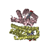
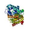
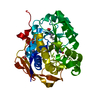
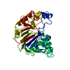
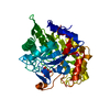
- PDB-4zva: Crystal structure of globin domain of the E. coli DosC - form I (... -
+ Open data

- Basic information
| Entry | Database: PDB / ID: 4zva | ||||||
|---|---|---|---|---|---|---|---|
| Title | Crystal structure of globin domain of the E. coli DosC - form I (ferric) | ||||||
 Components Components | Diguanylate cyclase DosC | ||||||
 Keywords Keywords | SIGNALING PROTEIN / oxygen sensing / diguanylate cyclase / cyclic-di-GMP / transferase | ||||||
| Function / homology |  Function and homology information Function and homology informationnegative regulation of bacterial-type flagellum-dependent cell motility / diguanylate cyclase / diguanylate cyclase activity / carbon monoxide binding / response to oxygen levels / cell adhesion involved in single-species biofilm formation / response to stress / oxygen binding / heme binding / GTP binding ...negative regulation of bacterial-type flagellum-dependent cell motility / diguanylate cyclase / diguanylate cyclase activity / carbon monoxide binding / response to oxygen levels / cell adhesion involved in single-species biofilm formation / response to stress / oxygen binding / heme binding / GTP binding / protein homodimerization activity / metal ion binding / plasma membrane Similarity search - Function | ||||||
| Biological species |  | ||||||
| Method |  X-RAY DIFFRACTION / SYNCHROTRON / SAD / Resolution: 2 Å X-RAY DIFFRACTION / SYNCHROTRON / SAD / Resolution: 2 Å | ||||||
 Authors Authors | Tarnawski, M. / Barends, T.R.M. / Schlichting, I. | ||||||
 Citation Citation | Journal: Acta Crystallogr.,Sect.D / Year: 2015 Title: Structural analysis of an oxygen-regulated diguanylate cyclase. Authors: Tarnawski, M. / Barends, T.R. / Schlichting, I. | ||||||
| History |
|
- Structure visualization
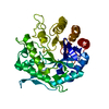
| Structure viewer | Molecule: MolmilJmol/JSmol |
|---|
- Downloads & links
-Download
| PDBx/mmCIF format | 4zva.cif.gz | 78.4 KB | Display | PDBx/mmCIF format |
|---|---|---|---|---|
| PDB format | pdb4zva.ent.gz | 58.2 KB | Display | PDB format |
| PDBx/mmJSON format | 4zva.json.gz | Tree view | PDBx/mmJSON format | |
| Others |  Other downloads Other downloads |
-Validation report
| Arichive directory | https://data.pdbj.org/pub/pdb/validation_reports/zv/4zvaftp://data.pdbj.org/pub/pdb/validation_reports/zv/4zva | HTTPS FTP |
|---|
-Related structure data
| Related structure data | 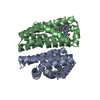 4zvbC 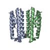 4zvcC 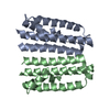 4zvdC 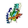 4zveC 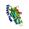 4zvfC 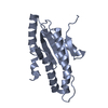 4zvgC 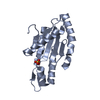 4zvhC C: citing same article ( |
|---|---|
| Similar structure data |

-Links
 PDBj
PDBj


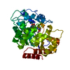
- Assembly
| Deposited unit | 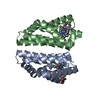
| |||||||||
|---|---|---|---|---|---|---|---|---|---|---|
| 1 |
| |||||||||
| Unit cell |
| |||||||||
| Components on special symmetry positions |
|
-Components
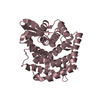
| #1: Protein | Mass: 18903.477 Da / Num. of mol.: 2 / Fragment: UNP residues 8-170 Source method: isolated from a genetically manipulated source Source: (gene. exp.) Gene: dosC, yddV, b1490, JW5241 / Production host: #2: Chemical |   Mass: 616.487 Da / Num. of mol.: 2 / Source method: obtained synthetically / Formula: C34H32FeN4O4 Mass: 616.487 Da / Num. of mol.: 2 / Source method: obtained synthetically / Formula: C34H32FeN4O4#3: Water | ChemComp-HOH / |  Mass: 18.015 Da / Num. of mol.: 137 / Source method: isolated from a natural source / Formula: H2O Mass: 18.015 Da / Num. of mol.: 137 / Source method: isolated from a natural source / Formula: H2O |
|---|
-Experimental details
-Experiment
| Experiment | Method: X-RAY DIFFRACTION |
|---|
- Sample preparation
| Crystal | Density Matthews: 2.76 Å3/Da / Density % sol: 55.38 % |
|---|---|
| Crystal grow | Temperature: 293 K / Method: vapor diffusion Details: 0.1 M tri-sodium citrate pH 5.6, 0.2 M ammonium acetate, 27% (w/v) PEG 4000 |
-Data collection
| Diffraction | Mean temperature: 100 K |
|---|---|
| Diffraction source | Source: SYNCHROTRON / Site: SLS  / Beamline: X10SA / Wavelength: 0.97627 Å / Beamline: X10SA / Wavelength: 0.97627 Å |
| Detector | Type: DECTRIS PILATUS 6M / Detector: PIXEL / Date: Oct 4, 2013 |
| Radiation | Monochromator: Si(111) / Protocol: SINGLE WAVELENGTH / Monochromatic (M) / Laue (L): M / Scattering type: x-ray |
| Radiation wavelength | Wavelength: 0.97627 Å / Relative weight: 1 |
| Reflection | Resolution: 2→50 Å / Num. obs: 29494 / % possible obs: 99.9 % / Redundancy: 10.7 % / Rmerge(I) obs: 0.05 / Net I/σ(I): 26.44 |
| Reflection shell | Resolution: 2→2.1 Å / Redundancy: 11 % / Rmerge(I) obs: 0.684 / Mean I/σ(I) obs: 3.52 / % possible all: 99.7 |
- Processing
| Software |
| ||||||||||||||||||||||||||||||||||||||||||||||||||||||||||||||||||||||||||||||||||||
|---|---|---|---|---|---|---|---|---|---|---|---|---|---|---|---|---|---|---|---|---|---|---|---|---|---|---|---|---|---|---|---|---|---|---|---|---|---|---|---|---|---|---|---|---|---|---|---|---|---|---|---|---|---|---|---|---|---|---|---|---|---|---|---|---|---|---|---|---|---|---|---|---|---|---|---|---|---|---|---|---|---|---|---|---|---|
| Refinement | Method to determine structure: SAD / Resolution: 2→48.705 Å / SU ML: 0.22 / Cross valid method: FREE R-VALUE / σ(F): 1.37 / Phase error: 26.99 / Stereochemistry target values: ML
| ||||||||||||||||||||||||||||||||||||||||||||||||||||||||||||||||||||||||||||||||||||
| Solvent computation | Shrinkage radii: 0.9 Å / VDW probe radii: 1.11 Å / Solvent model: FLAT BULK SOLVENT MODEL | ||||||||||||||||||||||||||||||||||||||||||||||||||||||||||||||||||||||||||||||||||||
| Refinement step | Cycle: LAST / Resolution: 2→48.705 Å
| ||||||||||||||||||||||||||||||||||||||||||||||||||||||||||||||||||||||||||||||||||||
| Refine LS restraints |
| ||||||||||||||||||||||||||||||||||||||||||||||||||||||||||||||||||||||||||||||||||||
| LS refinement shell |
|
