 Movie
Movie Controller

[English] 日本語
 Yorodumi
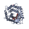
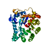
- PDB-6shd: Structure of the GH76A alpha-1,6-mannanase from Salegentibacter s... -
+ Open data

- Basic information
| Entry | Database: PDB / ID: 6shd | ||||||
|---|---|---|---|---|---|---|---|
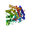
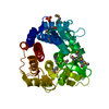
| Title | Structure of the GH76A alpha-1,6-mannanase from Salegentibacter sp. HEL1_6 | ||||||
 Components Components | Alpha-1,6-mannanase | ||||||
 Keywords Keywords | HYDROLASE / Mannanase / Glycoside hydrolase / GH76 / CAZyme / Mannan / Carbohydrate | ||||||
| Biological species |  Salegentibacter sp. Hel_I_6 (bacteria) Salegentibacter sp. Hel_I_6 (bacteria) | ||||||
| Method |  X-RAY DIFFRACTION / SYNCHROTRON / MOLECULAR REPLACEMENT / Resolution: 2 Å X-RAY DIFFRACTION / SYNCHROTRON / MOLECULAR REPLACEMENT / Resolution: 2 Å | ||||||
 Authors Authors | Hehemann, J.H. / Solanki, V. | ||||||
| Funding support |  Germany, 1items Germany, 1items
| ||||||
 Citation Citation | Journal: Isme J / Year: 2022 Title: Glycoside hydrolase from the GH76 family indicates that marine Salegentibacter sp. Hel_I_6 consumes alpha-mannan from fungi. Authors: Solanki, V. / Kruger, K. / Crawford, C.J. / Pardo-Vargas, A. / Danglad-Flores, J. / Hoang, K.L.M. / Klassen, L. / Abbott, D.W. / Seeberger, P.H. / Amann, R.I. / Teeling, H. / Hehemann, J.H. | ||||||
| History |
|
- Structure visualization
| Structure viewer | Molecule:  MolmilJmol/JSmol MolmilJmol/JSmol |
|---|
- Downloads & links
-Download
| PDBx/mmCIF format | 6shd.cif.gz | 590.2 KB | Display | PDBx/mmCIF format |
|---|---|---|---|---|
| PDB format | pdb6shd.ent.gz | 496.6 KB | Display | PDB format |
| PDBx/mmJSON format | 6shd.json.gz | Tree view | PDBx/mmJSON format | |
| Others |  Other downloads Other downloads |
-Validation report
| Arichive directory | https://data.pdbj.org/pub/pdb/validation_reports/sh/6shdftp://data.pdbj.org/pub/pdb/validation_reports/sh/6shd | HTTPS FTP |
|---|
-Related structure data
| Related structure data | 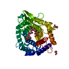 6shmC  6y8fC 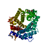 3k7xS S: Starting model for refinement C: citing same article ( |
|---|---|
| Similar structure data |


-Links
 PDBj
PDBj- Assembly
| Deposited unit | 
| ||||||||
|---|---|---|---|---|---|---|---|---|---|
| 1 | 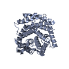
| ||||||||
| 2 | 
| ||||||||
| 3 | 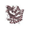
| ||||||||
| Unit cell |
|
-Components
| #1: Protein | Mass: 43747.512 Da / Num. of mol.: 3 Source method: isolated from a genetically manipulated source Source: (gene. exp.) Salegentibacter sp. Hel_I_6 (bacteria) / Gene: GH76A / Production host: #2: Water | ChemComp-HOH / |  Mass: 18.015 Da / Num. of mol.: 557 / Source method: isolated from a natural source / Formula: H2O Mass: 18.015 Da / Num. of mol.: 557 / Source method: isolated from a natural source / Formula: H2O |
|---|
-Experimental details
-Experiment
| Experiment | Method: X-RAY DIFFRACTION / Number of used crystals: 1 |
|---|
- Sample preparation
| Crystal | Density Matthews: 2.83 Å3/Da / Density % sol: 56.48 % |
|---|---|
| Crystal grow | Temperature: 289 K / Method: vapor diffusion, sitting drop / pH: 8 Details: 0.2 M Magnesium chloride hexahydrate 0.1 M Sodium acetate pH 5.0 20 % w/v PEG 6000 PH range: 7-8 |
-Data collection
| Diffraction | Mean temperature: 100 K / Serial crystal experiment: N | ||||||||||||||||||||||||||||||
|---|---|---|---|---|---|---|---|---|---|---|---|---|---|---|---|---|---|---|---|---|---|---|---|---|---|---|---|---|---|---|---|
| Diffraction source | Source: SYNCHROTRON / Site: PETRA III, DESY / Beamline: P11 / Wavelength: 0.9763 Å | ||||||||||||||||||||||||||||||
| Detector | Type: DECTRIS EIGER X 16M / Detector: PIXEL / Date: Oct 27, 2018 | ||||||||||||||||||||||||||||||
| Radiation | Protocol: SINGLE WAVELENGTH / Monochromatic (M) / Laue (L): M / Scattering type: x-ray | ||||||||||||||||||||||||||||||
| Radiation wavelength | Wavelength: 0.9763 Å / Relative weight: 1 | ||||||||||||||||||||||||||||||
| Reflection | Resolution: 2→92.02 Å / Num. obs: 87258 / % possible obs: 99.4 % / Redundancy: 6.6 % / CC1/2: 0.997 / Rmerge(I) obs: 0.1 / Rpim(I) all: 0.042 / Rrim(I) all: 0.108 / Net I/σ(I): 11.3 / Num. measured all: 574248 / Scaling rejects: 49 | ||||||||||||||||||||||||||||||
| Reflection shell | Diffraction-ID: 1
|
- Processing
| Software |
| ||||||||||||||||||||||||||||||||||||||||||||||||||||||||||||||||||||||||||||||||||||||||||
|---|---|---|---|---|---|---|---|---|---|---|---|---|---|---|---|---|---|---|---|---|---|---|---|---|---|---|---|---|---|---|---|---|---|---|---|---|---|---|---|---|---|---|---|---|---|---|---|---|---|---|---|---|---|---|---|---|---|---|---|---|---|---|---|---|---|---|---|---|---|---|---|---|---|---|---|---|---|---|---|---|---|---|---|---|---|---|---|---|---|---|---|
| Refinement | Method to determine structure: MOLECULAR REPLACEMENT Starting model: 3K7X Resolution: 2→55.091 Å / SU ML: 0.23 / Cross valid method: THROUGHOUT / σ(F): 1.33 / Phase error: 19.34
| ||||||||||||||||||||||||||||||||||||||||||||||||||||||||||||||||||||||||||||||||||||||||||
| Solvent computation | Shrinkage radii: 0.9 Å / VDW probe radii: 1.11 Å | ||||||||||||||||||||||||||||||||||||||||||||||||||||||||||||||||||||||||||||||||||||||||||
| Displacement parameters | Biso max: 85.62 Å2 / Biso mean: 39.0577 Å2 / Biso min: 10.76 Å2 | ||||||||||||||||||||||||||||||||||||||||||||||||||||||||||||||||||||||||||||||||||||||||||
| Refinement step | Cycle: final / Resolution: 2→55.091 Å
| ||||||||||||||||||||||||||||||||||||||||||||||||||||||||||||||||||||||||||||||||||||||||||
| LS refinement shell | Refine-ID: X-RAY DIFFRACTION / Rfactor Rfree error: 0
|
