 Movie
Movie Controller

[English] 日本語
 Yorodumi
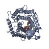
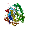
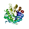
- PDB-3ipw: Crystal structure of hydrolase TatD family protein from Entamoeba... -
+ Open data

- Basic information
| Entry | Database: PDB / ID: 3ipw | ||||||
|---|---|---|---|---|---|---|---|
| Title | Crystal structure of hydrolase TatD family protein from Entamoeba histolytica | ||||||
 Components Components | Hydrolase TatD family protein | ||||||
 Keywords Keywords | HYDROLASE / NIAID / SSGCID / Seattle Structural Genomics Center for Infectious Disease / dysentery / liver abcess | ||||||
| Function / homology |  Function and homology information Function and homology information | ||||||
| Biological species |  Entamoeba histolytica HM-1:IMSS (eukaryote) Entamoeba histolytica HM-1:IMSS (eukaryote) | ||||||
| Method |  X-RAY DIFFRACTION / MOLECULAR REPLACEMENT / molecular replacement / Resolution: 1.95 Å X-RAY DIFFRACTION / MOLECULAR REPLACEMENT / molecular replacement / Resolution: 1.95 Å | ||||||
 Authors Authors | Seattle Structural Genomics Center for Infectious Disease (SSGCID) | ||||||
 Citation Citation | Journal: To be Published Title: Crystal structure of hydrolase TatD family protein from Entamoeba histolytica Authors: Edwards, T.E. / Statnekov, J.B. / Staker, B.L. / Seattle Structural Genomics Center for Infectious Disease (SSGCID) | ||||||
| History |
|
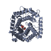
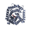
- Structure visualization
| Structure viewer | Molecule: MolmilJmol/JSmol |
|---|
- Downloads & links
-Download
| PDBx/mmCIF format | 3ipw.cif.gz | 78.3 KB | Display | PDBx/mmCIF format |
|---|---|---|---|---|
| PDB format | pdb3ipw.ent.gz | 56.1 KB | Display | PDB format |
| PDBx/mmJSON format | 3ipw.json.gz | Tree view | PDBx/mmJSON format | |
| Others |  Other downloads Other downloads |
-Validation report
| Arichive directory | https://data.pdbj.org/pub/pdb/validation_reports/ip/3ipwftp://data.pdbj.org/pub/pdb/validation_reports/ip/3ipw | HTTPS FTP |
|---|
-Related structure data
| Related structure data | 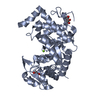 3e2vS S: Starting model for refinement |
|---|---|
| Similar structure data | |
| Other databases |


-Links
 PDBj
PDBj

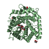
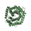
- Assembly
| Deposited unit | 
| ||||||||
|---|---|---|---|---|---|---|---|---|---|
| 1 |
| ||||||||
| Unit cell |
| ||||||||
| Details | Monomer |
-Components
| #1: Protein | Mass: 37512.719 Da / Num. of mol.: 1 Source method: isolated from a genetically manipulated source Source: (gene. exp.) Entamoeba histolytica HM-1:IMSS (eukaryote)Gene: EHI_119490 / Production host:  |
|---|---|
| #2: Chemical | ChemComp-GOL /   Mass: 92.094 Da / Num. of mol.: 1 / Source method: obtained synthetically / Formula: C3H8O3 Mass: 92.094 Da / Num. of mol.: 1 / Source method: obtained synthetically / Formula: C3H8O3 |
| #3: Water | ChemComp-HOH /  Mass: 18.015 Da / Num. of mol.: 157 / Source method: isolated from a natural source / Formula: H2O Mass: 18.015 Da / Num. of mol.: 157 / Source method: isolated from a natural source / Formula: H2O |
-Experimental details
-Experiment
| Experiment | Method: X-RAY DIFFRACTION / Number of used crystals: 1 |
|---|
- Sample preparation
| Crystal | Density Matthews: 2.09 Å3/Da / Density % sol: 41.29 % |
|---|---|
| Crystal grow | Temperature: 289 K / Method: vapor diffusion, sitting drop / pH: 7.5 Details: JCSG+ condition D1, 24% PEG 1500, 20% glycerol, 44.6 mg/mL protein, crystal tracking ID 202317d1, pH 7.5, VAPOR DIFFUSION, SITTING DROP, temperature 289K |
-Data collection
| Diffraction | Mean temperature: 100 K |
|---|---|
| Diffraction source | Source: ROTATING ANODE / Type: RIGAKU FR-E+ SUPERBRIGHT / Wavelength: 1.5418 Å |
| Detector | Type: RIGAKU SATURN 944+ / Detector: CCD / Date: Aug 7, 2009 |
| Radiation | Protocol: SINGLE WAVELENGTH / Monochromatic (M) / Laue (L): M / Scattering type: x-ray |
| Radiation wavelength | Wavelength: 1.5418 Å / Relative weight: 1 |
| Reflection | Resolution: 1.95→50 Å / Num. obs: 23615 / % possible obs: 98.8 % / Redundancy: 6 % / Rmerge(I) obs: 0.073 / Χ2: 1.034 / Net I/σ(I): 21.8 |
| Reflection shell | Resolution: 1.95→2.02 Å / Redundancy: 3.7 % / Rmerge(I) obs: 0.47 / Mean I/σ(I) obs: 2.45 / Num. unique all: 2166 / Χ2: 0.971 / % possible all: 93.2 |
-Phasing
| Phasing | Method: molecular replacement | |||||||||
|---|---|---|---|---|---|---|---|---|---|---|
| Phasing MR | Rfactor: 57.35 / Model details: Phaser MODE: MR_AUTO
|
- Processing
| Software |
| |||||||||||||||||||||||||||||||||||||||||||||||||||||||||||||||||
|---|---|---|---|---|---|---|---|---|---|---|---|---|---|---|---|---|---|---|---|---|---|---|---|---|---|---|---|---|---|---|---|---|---|---|---|---|---|---|---|---|---|---|---|---|---|---|---|---|---|---|---|---|---|---|---|---|---|---|---|---|---|---|---|---|---|---|
| Refinement | Method to determine structure: MOLECULAR REPLACEMENT Starting model: 3e2v clipped to contain only homologous sections Resolution: 1.95→50 Å / Cor.coef. Fo:Fc: 0.953 / Cor.coef. Fo:Fc free: 0.93 / WRfactor Rfree: 0.22 / WRfactor Rwork: 0.173 / Occupancy max: 1 / Occupancy min: 0.5 / FOM work R set: 0.83 / SU B: 3.68 / SU ML: 0.107 / SU R Cruickshank DPI: 0.176 / SU Rfree: 0.163 / Cross valid method: THROUGHOUT / σ(F): 0 / ESU R: 0.176 / ESU R Free: 0.163 / Stereochemistry target values: MAXIMUM LIKELIHOOD Details: HYDROGENS HAVE BEEN ADDED IN THE RIDING POSITIONS U VALUES : REFINED INDIVIDUALLY
| |||||||||||||||||||||||||||||||||||||||||||||||||||||||||||||||||
| Solvent computation | Ion probe radii: 0.8 Å / Shrinkage radii: 0.8 Å / VDW probe radii: 1.4 Å / Solvent model: MASK | |||||||||||||||||||||||||||||||||||||||||||||||||||||||||||||||||
| Displacement parameters | Biso max: 56.28 Å2 / Biso mean: 25.689 Å2 / Biso min: 10.4 Å2
| |||||||||||||||||||||||||||||||||||||||||||||||||||||||||||||||||
| Refinement step | Cycle: LAST / Resolution: 1.95→50 Å
| |||||||||||||||||||||||||||||||||||||||||||||||||||||||||||||||||
| Refine LS restraints |
| |||||||||||||||||||||||||||||||||||||||||||||||||||||||||||||||||
| LS refinement shell | Resolution: 1.951→2.001 Å / Total num. of bins used: 20
|
