 Movie
Movie Controller

[English] 日本語
 Yorodumi
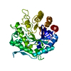
- PDB-6shm: An inactive (D136A and D137A) variant of alpha-1,6-mannanase, GH7... -
+ Open data

- Basic information
| Entry | Database: PDB / ID: 6shm | ||||||
|---|---|---|---|---|---|---|---|
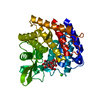
| Title | An inactive (D136A and D137A) variant of alpha-1,6-mannanase, GH76A of Salegentibacter sp. HEL1_6 in complex with alpha-1,6-mannotetrose | ||||||
 Components Components | Mutant alpha-1,6-mannanase GH76A | ||||||
 Keywords Keywords | HYDROLASE / Glycoside hydrolase / Mannanase / mannotetrose / GH76 / CAZyme / polysaccharide | ||||||
| Biological species |  Salegentibacter sp. Hel_I_6 (bacteria) Salegentibacter sp. Hel_I_6 (bacteria) | ||||||
| Method |  X-RAY DIFFRACTION / SYNCHROTRON / MOLECULAR REPLACEMENT / Resolution: 1.9 Å X-RAY DIFFRACTION / SYNCHROTRON / MOLECULAR REPLACEMENT / Resolution: 1.9 Å | ||||||
 Authors Authors | Hehemann, J.H. / Solanki, V. | ||||||
| Funding support |  Germany, 1items Germany, 1items
| ||||||
 Citation Citation | Journal: Isme J / Year: 2022 Title: Glycoside hydrolase from the GH76 family indicates that marine Salegentibacter sp. Hel_I_6 consumes alpha-mannan from fungi. Authors: Solanki, V. / Kruger, K. / Crawford, C.J. / Pardo-Vargas, A. / Danglad-Flores, J. / Hoang, K.L.M. / Klassen, L. / Abbott, D.W. / Seeberger, P.H. / Amann, R.I. / Teeling, H. / Hehemann, J.H. | ||||||
| History |
|
- Structure visualization
| Structure viewer | Molecule:  MolmilJmol/JSmol MolmilJmol/JSmol |
|---|
- Downloads & links
-Download
| PDBx/mmCIF format | 6shm.cif.gz | 165.6 KB | Display | PDBx/mmCIF format |
|---|---|---|---|---|
| PDB format | pdb6shm.ent.gz | 128.5 KB | Display | PDB format |
| PDBx/mmJSON format | 6shm.json.gz | Tree view | PDBx/mmJSON format | |
| Others |  Other downloads Other downloads |
-Validation report
| Arichive directory | https://data.pdbj.org/pub/pdb/validation_reports/sh/6shmftp://data.pdbj.org/pub/pdb/validation_reports/sh/6shm | HTTPS FTP |
|---|
-Related structure data
| Related structure data |  6shdSC  6y8fC S: Starting model for refinement C: citing same article ( |
|---|---|
| Similar structure data |


-Links
 PDBj
PDBj- Assembly
| Deposited unit | 
| ||||||||
|---|---|---|---|---|---|---|---|---|---|
| 1 |
| ||||||||
| Unit cell |
|
-Components
| #1: Protein | Mass: 43790.688 Da / Num. of mol.: 1 / Mutation: D136A and D137A Source method: isolated from a genetically manipulated source Details: WP_051935954.1 / Source: (gene. exp.) Salegentibacter sp. Hel_I_6 (bacteria) / Gene: GH76A / Plasmid: pET28a / Production host: |
|---|---|
| #2: Polysaccharide | alpha-D-mannopyranose-(1-6)-alpha-D-mannopyranose-(1-6)-alpha-D-mannopyranose-(1-6)-alpha-D-mannopyranose Source method: isolated from a genetically manipulated source |
| #3: Chemical | ChemComp-MES /   Mass: 195.237 Da / Num. of mol.: 1 / Source method: obtained synthetically / Formula: C6H13NO4S / Comment: pH buffer*YM Mass: 195.237 Da / Num. of mol.: 1 / Source method: obtained synthetically / Formula: C6H13NO4S / Comment: pH buffer*YM |
| #4: Chemical | ChemComp-GOL /   Mass: 92.094 Da / Num. of mol.: 1 / Source method: obtained synthetically / Formula: C3H8O3 Mass: 92.094 Da / Num. of mol.: 1 / Source method: obtained synthetically / Formula: C3H8O3 |
| #5: Water | ChemComp-HOH /  Mass: 18.015 Da / Num. of mol.: 351 / Source method: isolated from a natural source / Formula: H2O Mass: 18.015 Da / Num. of mol.: 351 / Source method: isolated from a natural source / Formula: H2O |
| Has ligand of interest | Y |
-Experimental details
-Experiment
| Experiment | Method: X-RAY DIFFRACTION / Number of used crystals: 1 |
|---|
- Sample preparation
| Crystal | Density Matthews: 1.98 Å3/Da / Density % sol: 37.76 % |
|---|---|
| Crystal grow | Temperature: 289 K / Method: vapor diffusion, sitting drop / pH: 8 Details: 1.6 M Tri-sodium citrate 10 % Glycerol 0.1 M MES monohydrate pH 6.5 PH range: 6-8 |
-Data collection
| Diffraction | Mean temperature: 100 K / Serial crystal experiment: N | ||||||||||||||||||||||||||||||
|---|---|---|---|---|---|---|---|---|---|---|---|---|---|---|---|---|---|---|---|---|---|---|---|---|---|---|---|---|---|---|---|
| Diffraction source | Source: SYNCHROTRON / Site: PETRA III, DESY / Beamline: P11 / Wavelength: 1.0332 Å | ||||||||||||||||||||||||||||||
| Detector | Type: DECTRIS PILATUS 6M-F / Detector: PIXEL / Date: May 24, 2019 | ||||||||||||||||||||||||||||||
| Radiation | Protocol: SINGLE WAVELENGTH / Monochromatic (M) / Laue (L): M / Scattering type: x-ray | ||||||||||||||||||||||||||||||
| Radiation wavelength | Wavelength: 1.0332 Å / Relative weight: 1 | ||||||||||||||||||||||||||||||
| Reflection | Resolution: 1.9→63.48 Å / Num. obs: 24400 / % possible obs: 99.2 % / Redundancy: 9.3 % / CC1/2: 0.279 / Rmerge(I) obs: 5.441 / Rpim(I) all: 2.333 / Rrim(I) all: 0.5969 / Net I/σ(I): 7.3 | ||||||||||||||||||||||||||||||
| Reflection shell | Diffraction-ID: 1
|
- Processing
| Software |
| |||||||||||||||||||||||||||||||||||||||||||||||||||||||||||||||||
|---|---|---|---|---|---|---|---|---|---|---|---|---|---|---|---|---|---|---|---|---|---|---|---|---|---|---|---|---|---|---|---|---|---|---|---|---|---|---|---|---|---|---|---|---|---|---|---|---|---|---|---|---|---|---|---|---|---|---|---|---|---|---|---|---|---|---|
| Refinement | Method to determine structure: MOLECULAR REPLACEMENT Starting model: 6SHD Resolution: 1.9→47.51 Å / Cor.coef. Fo:Fc: 0.968 / Cor.coef. Fo:Fc free: 0.943 / SU B: 6.527 / SU ML: 0.085 / Cross valid method: THROUGHOUT / σ(F): 0 / ESU R Free: 0.122 Details: HYDROGENS HAVE BEEN ADDED IN THE RIDING POSITIONS U VALUES : REFINED INDIVIDUALLY
| |||||||||||||||||||||||||||||||||||||||||||||||||||||||||||||||||
| Solvent computation | Ion probe radii: 0.8 Å / Shrinkage radii: 0.8 Å / VDW probe radii: 1.2 Å | |||||||||||||||||||||||||||||||||||||||||||||||||||||||||||||||||
| Displacement parameters | Biso max: 71.61 Å2 / Biso mean: 12.801 Å2 / Biso min: 2.14 Å2
| |||||||||||||||||||||||||||||||||||||||||||||||||||||||||||||||||
| Refinement step | Cycle: final / Resolution: 1.9→47.51 Å
| |||||||||||||||||||||||||||||||||||||||||||||||||||||||||||||||||
| Refine LS restraints |
| |||||||||||||||||||||||||||||||||||||||||||||||||||||||||||||||||
| LS refinement shell | Resolution: 1.9→1.949 Å / Rfactor Rfree error: 0 / Total num. of bins used: 20
|
