 Movie
Movie Controller

[English] 日本語
 Yorodumi
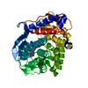
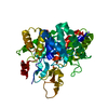
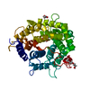
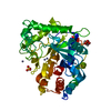
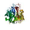
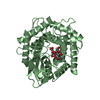
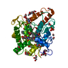
- PDB-2d5j: Unsaturated Glucuronyl Hydrolase Triggers Hydration of Vinyl Ethe... -
+ Open data

- Basic information
| Entry | Database: PDB / ID: 2d5j | ||||||
|---|---|---|---|---|---|---|---|
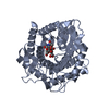
| Title | Unsaturated Glucuronyl Hydrolase Triggers Hydration of Vinyl Ether Group but not of Glycosidic Bond | ||||||
 Components Components | unsaturated glucuronyl hydrolase | ||||||
 Keywords Keywords | HYDROLASE / alpha/alpha barrel / unsaturated glucuronyl hydrolase | ||||||
| Function / homology |  Function and homology information Function and homology informationgellan tetrasaccharide unsaturated glucuronosyl hydrolase / chondroitin hydrolase activity / polysaccharide catabolic process / cytoplasm Similarity search - Function | ||||||
| Biological species |  | ||||||
| Method |  X-RAY DIFFRACTION / SYNCHROTRON / MOLECULAR REPLACEMENT / Resolution: 1.6 Å X-RAY DIFFRACTION / SYNCHROTRON / MOLECULAR REPLACEMENT / Resolution: 1.6 Å | ||||||
 Authors Authors | Itoh, T. / Hashimoto, W. / Mikami, B. / Murata, K. | ||||||
 Citation Citation | Journal: J.Biol.Chem. / Year: 2006 Title: Crystal Structure of Unsaturated Glucuronyl Hydrolase Complexed with Substrate: MOLECULAR INSIGHTS INTO ITS CATALYTIC REACTION MECHANISM Authors: Itoh, T. / Hashimoto, W. / Mikami, B. / Murata, K. | ||||||
| History |
|
- Structure visualization
| Structure viewer | Molecule: MolmilJmol/JSmol |
|---|
- Downloads & links
-Download
| PDBx/mmCIF format | 2d5j.cif.gz | 177.1 KB | Display | PDBx/mmCIF format |
|---|---|---|---|---|
| PDB format | pdb2d5j.ent.gz | 141.6 KB | Display | PDB format |
| PDBx/mmJSON format | 2d5j.json.gz | Tree view | PDBx/mmJSON format | |
| Others |  Other downloads Other downloads |
-Validation report
| Arichive directory | https://data.pdbj.org/pub/pdb/validation_reports/d5/2d5jftp://data.pdbj.org/pub/pdb/validation_reports/d5/2d5j | HTTPS FTP |
|---|
-Related structure data


-Links
 PDBj
PDBj
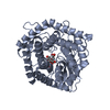
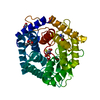
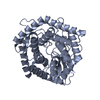
- Assembly
| Deposited unit | 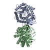
| ||||||||
|---|---|---|---|---|---|---|---|---|---|
| 1 | 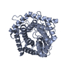
| ||||||||
| 2 | 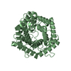
| ||||||||
| Unit cell |
|
-Components
| #1: Protein | Mass: 42914.352 Da / Num. of mol.: 2 Source method: isolated from a genetically manipulated source Source: (gene. exp.) References: UniProt: Q9RC92, Hydrolases; Glycosylases; Glycosidases, i.e. enzymes that hydrolyse O- and S-glycosyl compounds #2: Water | ChemComp-HOH / |  Mass: 18.015 Da / Num. of mol.: 835 / Source method: isolated from a natural source / Formula: H2O Mass: 18.015 Da / Num. of mol.: 835 / Source method: isolated from a natural source / Formula: H2O |
|---|
-Experimental details
-Experiment
| Experiment | Method: X-RAY DIFFRACTION / Number of used crystals: 1 |
|---|
- Sample preparation
| Crystal | Density Matthews: 2.33 Å3/Da / Density % sol: 47.18 % |
|---|---|
| Crystal grow | Temperature: 293 K / Method: vapor diffusion, hanging drop / pH: 8.6 Details: 30% PEG10000, 0.15M Tris pH 8.6, VAPOR DIFFUSION, HANGING DROP, temperature 293K |
-Data collection
| Diffraction | Mean temperature: 100 K |
|---|---|
| Diffraction source | Source: SYNCHROTRON / Site: SPring-8  / Beamline: BL38B1 / Wavelength: 1 Å / Beamline: BL38B1 / Wavelength: 1 Å |
| Detector | Type: RIGAKU JUPITER 210 / Detector: CCD / Date: Sep 27, 2005 |
| Radiation | Protocol: SINGLE WAVELENGTH / Monochromatic (M) / Laue (L): M / Scattering type: x-ray |
| Radiation wavelength | Wavelength: 1 Å / Relative weight: 1 |
| Reflection | Resolution: 1.6→50 Å / Num. obs: 93414 / % possible obs: 87.7 % / Redundancy: 4.3 % / Biso Wilson estimate: 15.9 Å2 / Rmerge(I) obs: 0.079 |
| Reflection shell | Resolution: 1.6→1.66 Å / Redundancy: 3.3 % / Rmerge(I) obs: 0.276 / % possible all: 96.1 |
- Processing
| Software |
| ||||||||||||||||||||||||||||||||||||||||||||||||||||||||||||||||||||||||||||||||
|---|---|---|---|---|---|---|---|---|---|---|---|---|---|---|---|---|---|---|---|---|---|---|---|---|---|---|---|---|---|---|---|---|---|---|---|---|---|---|---|---|---|---|---|---|---|---|---|---|---|---|---|---|---|---|---|---|---|---|---|---|---|---|---|---|---|---|---|---|---|---|---|---|---|---|---|---|---|---|---|---|---|
| Refinement | Method to determine structure: MOLECULAR REPLACEMENT / Resolution: 1.6→9.99 Å / Rfactor Rfree error: 0.002 / Data cutoff high absF: 1726790.34 / Data cutoff low absF: 0 / Isotropic thermal model: RESTRAINED / Cross valid method: THROUGHOUT / σ(F): 0 / Stereochemistry target values: Engh & Huber
| ||||||||||||||||||||||||||||||||||||||||||||||||||||||||||||||||||||||||||||||||
| Solvent computation | Solvent model: FLAT MODEL / Bsol: 65.697 Å2 / ksol: 0.451516 e/Å3 | ||||||||||||||||||||||||||||||||||||||||||||||||||||||||||||||||||||||||||||||||
| Displacement parameters | Biso mean: 16.8 Å2
| ||||||||||||||||||||||||||||||||||||||||||||||||||||||||||||||||||||||||||||||||
| Refine analyze |
| ||||||||||||||||||||||||||||||||||||||||||||||||||||||||||||||||||||||||||||||||
| Refinement step | Cycle: LAST / Resolution: 1.6→9.99 Å
| ||||||||||||||||||||||||||||||||||||||||||||||||||||||||||||||||||||||||||||||||
| Refine LS restraints |
| ||||||||||||||||||||||||||||||||||||||||||||||||||||||||||||||||||||||||||||||||
| LS refinement shell | Resolution: 1.6→1.7 Å / Rfactor Rfree error: 0.007 / Total num. of bins used: 6
| ||||||||||||||||||||||||||||||||||||||||||||||||||||||||||||||||||||||||||||||||
| Xplor file |
|
